Wednesday, February 13, 2013

Classification of Tracheo-Esophageal Fistula


This classification is anatomically based.

The commonest is still the atretic esophagus with distal fistula as on the left. The middle figure is the isolated Tracheo-esophageal fistula or also known as the 'H' type. Other's are not as common.

Surgical management differs according to anatomical variations.

Other Topics:
- Diagnosis
- Management
- Associated anomalies and VACTERL

Tuesday, February 5, 2013

Kasai Procedure


In 1959, Morio Kasai, a Japanese surgeon discovered a breakthrough in the treatment of biliary atresia which had a poor survival rate before. The procedure which carries his name, although modified in various forms, is still performed today for patient's with biliary atresia.

Procedure


The principle of the procedure is the resect the atretic parts of the extrahepatic biliary tree, dissect the porta hepatis of the liver to free up patent biliary ductules and anastamose with the intestine in a Roux-en-Y fashion (portoenterostomy).

A right upper abdominal incision is made (though from my observation here, the roof top incision is popular). Other anomalies should be sought out and excluded (polysplenia or asplenia, malrotation, preduodenal portal vein, interrupted vena cava).

Before dissection of the portal plate (porta hepatis at the liver surface), examination of the extrahapatic biliary tract is made. If the gallbladder is atretic and without lumen, the surgery may then proceed with the Kasai. If not, an on-table cholangiogram must be performed to delineate the extend of atresia or if atresia exist at all. Failure to outline patent intrahepatic and extrahaptic biliary structures justifies the need to proceed with portoenterostomy. 

The hepatoduodenal ligament (the free edge of the lesser omentum) is opened up to identify the structures of the bile duct and is dissected away from the hepatic arteries. Dissection is continued proximally towards the liver, the gallbladder also dissected. The end-point of dissection is after the bifurcation of the portal vein. A fibrous cone and portal plate should be seen here.

At this stage, the fibrous cone should be transected with knife or scissors. This is the most crucial step to the success of surgery, too deep a cut will cause injury to the liver and cause subsequent scarring and obliteration of the biliary ductules. Too superficial, patent biliary ductules are not exposed enough for drainage. The use of diathermy should be discouraged as this may damage the fine ductules that is essential to the success of the surgery. Care is taken to not injure the portal vein to avoid bleeding.

The proximal jejenum, about 10 cm from the Ligament of Trietze is identified and transected. The distal end of the transection is anatamosed with the liver with 6/0 absorbable sutures brought through the avascular portion of the mesocolon. End-to-side jejunojenunostomy is created at about 50 - 60 cm from the transected part of the jejunum. The defect in the mesocolon is closed by anchoring the roux limb. This is done to prevent internal herniation and keep the limb without tension.

Placement of a suction drain is recommended by some near the portoenterostomy site.

Post-Op

Return of 'color' to stool is an indication of success which usually occur after 10 - 14 days. However some will still progress to liver failure despite initial cholic stool, and some remain with acholic stool.

NG tube is continued until about 48 hours post-op.

Recommended medical regimen includes:
Choleretic: Ursidol : 10 - 15 mg/kg/dose bd
Trimethoprim-sulfamethoxazole (Bactrim) : 2.5 mg/kg/day
Vitamin ADEK
Prednisolone : 2mg/mg/day to be tapered down over 6 weeks.

Factors to Success

Age of surgery seem to be an important factor. Most literature report success if the procedure is performed at 70 to 90 days old. This however does not contraindicate surgery in older children.

Patent gallbladder and fibrous cones are also indicators to better prognosis. So does the diameter of ductules.

Complications

Cholangitis:
An important complication that may lead to the next devastating complication, portal hypertension. Prevention is best done by performing an adequate roux surgery, as well as antibiotics prophylaxis during surgery and post-operatively. The use of steroids also helps in reducing inflammation that may also prevent scarring of the liver although presenting evidence is controversial (not discussed here). Otherwise, treatment is best done with broad-spectrum antibiotics that covers anaerobes as well.

Portal Hypertension:
The usual triad may form including ascites, hypersplenism with associated thrmbocytopenia, and the feared esophageal varices that may lead to bleeding. Liver transplant is indicated if this complication is severe.

Intrahepatic biliary cavities of cyst:
May develop within the liver that may contribute to recurrent cholangitis. Larger ones may be drained percutaneously.

Others:
Wound dehiscence
Internal herniation through the mesocolon defect.
Anastamotic leak
Intussuception at the foot of the roux.

Malignancies:
Cirrhotic liver may lead to hepatocellular carcinoma and hepatoblastoma which all have been reported in patient's with biliary atresia.


References:

1. Coran, Arnold G., et al., Pediatric Surgery, 7th Ed., Elsevier

2. Wildhaber, Barbara E., Biliary Atresia: 50 Years After the First Kasai, Review Article, ISRN Surgery (2012).  Must read. Contains other articles on modified Kasai procedures and others.


Future Topics:

Biliary atresia
Liver Transplant
Complications of biliary atresia (in details)

Monday, February 4, 2013

Use of Negative Pressure Wound therapy for Abdominal Wounds in Neonates and Infants: Critical Appraisal

Use of Negative Pressure Wound Therapy (NPWT) for Abdominal Wounds in Neonates and Infants, Journal of Pediatric Surgery (2012) 47, 1555-1559, Stoffan, Alexander P., et al.

Summary of Article:

The article described the use of NPWT for the past 10 years in Children's Hospital Boston and its outcome. There is no attempts in studying it effectiveness or in comparison to other methods of wound closure or treatment. Only 2 cases developed fistula after initiation of NPWT. Most of the other cases have already developed stoma and fistula before NPWT was applied. 6 died from the cohort but none were related to NPWT.

My 2 cents:

While the article describes the experience of NPWT in their institution, there is a lack useful description to describe the efficacy of NPWT in this age group which is understandable as the article does not intend to report on the subject.

The number of subjects are also too few (18) to be of much benefit.

The wide difference in diagnosis of wounds used for NPWT also makes it difficult to tell if the diagnosis makes a difference to the success of NPWT although some of their results sounds convincing.

Discussion:

Wounds and chronic wounds are a hassle to manage, especially post-operatively. Chronic wounds is a frustrating complications for all parties, including

While not exactly new, its use it being increasingly used in the treatment of especially chronic wounds although any wounds as long as it is not contraindicated, may be treated with this method. In my work in Queen Elizabeth Hospital in Kota Kinabalu, Sabah, its use is increasing especially in the orthopedic's department and general surgery and plastic surgery departments.

The principle to NPWT is that the negative pressure when applied in an air tight seal on a wound, will draw fluids from the wound decreasing edema and encouraging wound healing. It is also said to increase blood flow to the wound which is an important factor in the healing of the wound.

While it is being used in the treatment of wound in adults, its use in the pediatric age group, especially the infant and neonates are also increasing. However, there is a lack of evidence from literature to its efficacy. This is mostly due to the heterogenicity of diagnosis and types of wounds that makes controlled random clinical trials difficult without bias. Despite this, it cannot be denied that NPWT remains an important tool that needs to be utilized.

More research needs to be done surrounding the subject. Wound treatment in the infant and neonate age group may certainly benefit from this modality of treatment. Staff needs to be educated and trained in the application of this technique as it may provide a different alternative when traditional methods of wound care fails.


References:

1. Stoffan, Alexander P., et al., Use of Negative Pressure Wound Therapy (NPWT) for Abdominal Wounds in Neonates and Infants, Journal of Pediatric Surgery (2012) 47, 1555-1559

2. Gestring M, et al., Negative Wound Pressure Therapy, UpToDate Article (2012)

3. Gregor S., et al., Negative Pressure Wound Therapy: A Vacuum of Evidence? Arch Surg 2008

Future Topics:
  • Wound closure methods
  • Wound healing in children

Friday, February 1, 2013

Sistrunk Procedure

Walter Ellis Sistrunk described the surgery for the excision of the thyroglossal duct cyst (TGDC) in 1920.

The principle of the surgery is to remove the cyst along with its tract and the tissues surrounding it including part of the hyoid bone to reduce rate of recurrence of the cyst.

The embryological pathway for the descent of the thyroid gland starting from the foramen caecum, crossing the hyoid bone.


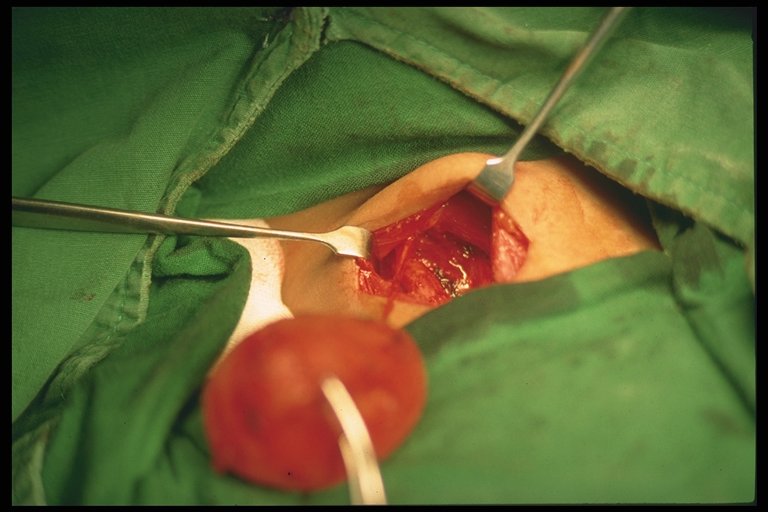
The patient is placed in supine position. A transverse cervical incision is made along the hyoid bone. The cyst and tract is mobilized. A portion of the hyoid bone, about 1 cm each side form the midline is excised after releasing the hyoglossus and mylohyoid muscles. Tissues surrounding the ducts are excised up to the foramen caecum of the tongue. No attempts are made at separating the ducts from the tissue as Sistrunk noted  that the duct are friable and easily broken. The defects are then approximated including the cut hyoid bones.


Cyst with parts of the ducts proximal to it.

On the right is the excised TGDC with a part of the excised hyoid bone and proximal tissues on the left.

Recurrence is about 10% after surgery. Wider excision is recommended for a recurrent cyst. Infected cysts are best drained or treated with antibiotics before proceding with surgery.


References:

1. Sistrunk, W. E., The Surgical Treatment of Cyst of the Thyroglossal Tract, Reprinted from Ann Surg 1920.

2. Coran, Arnold G., et al., Pediatric Surgery, 7th Ed., Elsevier

3. Pictures from various sites as linked.

Other Topics:

- Neck masses in children
- Embryology of the brachial arches

Friday, January 25, 2013

Twin-Twin Transfusion Syndrome: Clinical Features


Twin to twin transfusion syndrome (TTTS) is a condition where there is an over-perfusion in one twin and under-perfusion in the other. Although more common in monochorionic twins, it has been reported to occur in diamniotic twins.

Clinical Features and Diagnosis:

Most important is the discrepancy in amniotic fluid volume.
1 twin would be oligohydramnios ( < 2cm deep vertical pool )
1 twin would be polyhydramnios ( > 8cm deep vertical pool)

WEIGHT DISCREPANCY IS NOT THE MAIN CRITERIA FOR DIAGNOSIS!
In some cases, there are no significant weight discrepancies.
If there are, the discordance should be > 15-20%

"Stuck twin appearance"


Anhydromnios fetus appear as if there are no separating membrane.

Discrepancies in size of umbilical cords.

Presence of hydrops or cardiac dysfunction in recipient twin.

Abnormal umbilical artery doppler in donor fetus.

Other features of monochorionic twins:
  • Single placenta
  • Gender concordance
Fetal Blood Sampling:
- Lower hematocrit level in donors
- Difference in hemoglobin levels.

Staging:

Quintero staging uses simple concept of staging involving presence/absence of oligo or polyhydromnios, bladder, abnormal dopple, hydrops fetalis, or fetal death.

A table can be seen in this Emedicine website.

References:

Creasy, Robert K., et al., Maternal-Fetal Medicine, Principles and Practice, 5th Ed.

Other Topics To Discuss:

- Management of TTTS
- Monochorionic twins
- Conjoint twins.


Thursday, January 24, 2013

Renal Trauma: Short Notes

Genitourinary tract traumas are common in the pediatric age group consisting of injuries to the kidneys, ureter, bladder, and genitalia. 50% of genitourinary tract trauma involves the kidney. In this short notes I will talk about renal trauma. As in most solid organ traumas, it can be managed non-operatively in the majority of patients.

Epidemiology
  • Injuries are classified into blunt and penetrating injuries.
  • MVAs make up about 60% of the cause of renal injuries, followed by falls (20%) and sports injuries (10%).
  • Sudden decelerations with flexion-extension movement in seat-belt is a known mechanism of injury
  • In the USA, penetrating injuries are caused mostly by gunshot wounds (86%) followed by stab wounds.
  • Associated injuries include liver, spleen and bowel injury. These injuries most be sought for and ruled out.
Kidneys in Children

Certain features and differences in the child's kidneys make them more susceptible to injury compared to adults.
  • Kidneys are relative larger compared to the size of the child's body.
  • Position is much lower as well in the abdomen.
  • Less protected due to decreased perirenal fat.
  • Renal capsule and Gerota's fascia less well-developed.
  • More mobile kidneys at the pedicle make it more susceptible to deceleration injuries.
  • Retainment of lobulations increase risk of parenchymal distruption.
  • Weaker abdominal wall muscles and poorly ossified rib cages make renal injury more likely.
  • Congenital anomalies increases risk ie. hydronephrosis, horseshoe kidney, polycystic kidneys, renal tumors.
Clinical Features
  • Abdominal pain and flank pain.
  • Flank echymosis
  • Hematuria (absence does not exclude underlying injury)
  • Associated injuries include lower ribs and lumbar vertabrae fracture.
  • A lot of children are also asymptomatic
Investigations
  • FBC, Renal function
  • Urinalysis (controversy in further radiological imaging if microscopic hematuria is present. Request for imaging must be correlated with the whole clinical picture).
Imaging
  • Contrasted abdominal CT-scan is standard for evaluation.
  • Intravenous pyelography useful in unstable patient prior to op to determine functioning of 2 kidneys, extent of urinary extravasation, pedicle injuries.
  • Ultrasound not sensitive in detecting parenchymal injuries. Useful for follow-ups to exclude urinomas, expanding hematomas, abscess, pseudoaneurysm.
Grading of Injury

From: Simple Medicine website, http://simple-med.blogspot.com/


 American Association for the Surgery of Trauma (AAST) Grading for Renal Injury

Grade I: Subcapsular, nonexpanding hematoma. Microscopic or gross hematuria with normal urologic studies in contusions.

Grade II: < 1 cm parenchymal depth of cortex laceration without urinary extravasation. Non-expanding hematoma confined to renal retroperitoneum.

Grade III: > 1 cm parenchymal depth of cortex laceration without urinary extravasation.

Grade IV: Laceration extending up to cortex, medulla and collecting system. Renal artery or vein injury with contained hemorrhage.

Grade V: Complete shattered kidney. Avulsion of renal hilum that devascularizes the kidney.

*Grading to standardize description for research and collection purposes.
**Generally the lower the grade I - III, the less likely need for operative intevention provided patient is stable.
***This grading is developed for adults that although may apply for children, but management should be based on case to case basis.

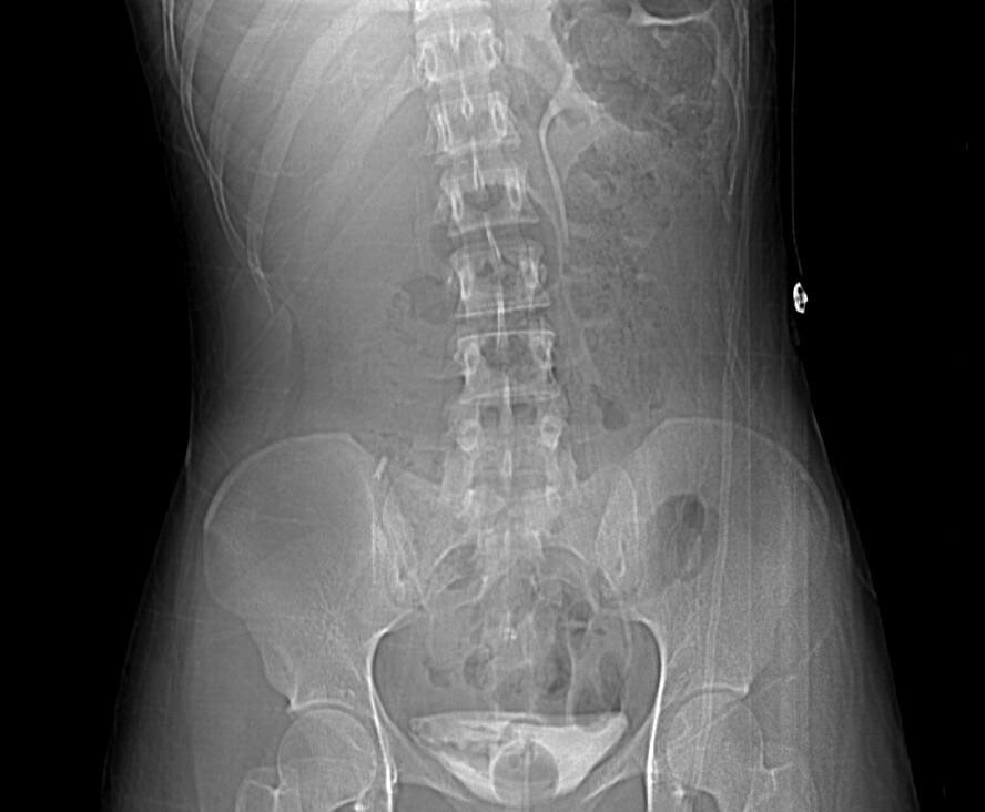
The following pictures are not only from pediatric cases.

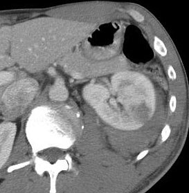
Grade III renal injury. Hypoechoic lesion in the left kidney after an MVA.

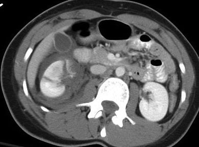
Grade V renal injury. Note the lacerations through and through the left kidney with surrounding hematoma.
Management
  • As mentioned, most (~98%) require only bed rest and observation in stable patients, even in grade IV and V injuries.
  • However, falling blood counts (Hb), persistent gross hematuria, hemodynamic instability and multiple transfusion requirements may indicate ongoing bleeding.
  • Arteriography and embolization to be considered in selective cases. 
  • Unstable patients may need explorative laparotomy.
  • Nephrectomy to be considered in extensive unsalvagable injuries, especially in hemodynamically unstable patients. 
  • Preservation of vessels attempted in cases of solitary kidneys or bilateral non-functioning kidneys.
Complications of Non-operative Management
  • Ongoing bleeding.
  • Urinary extravasation, may present as ileus, abdominal flank mass or discomfort. Urinomas, most resolve spontaneously.
  • Perinephric abscess.
  • Hydronephrosis
  • Arterio-venous fistula
  • Pseudoaneurysm
  • Pyelonephritis
  • Renal calculi
  • Hypertension, may be delayed.
Follow-up
  • Most have normal renal function and without hypertension.
  • The above complications need to be sought out.
  • Further imaging (ultrasonography to reduce radiation exposure) may be required if complications develops. 
Challenge:

Describe the images:


References:

Coran, Arnold G., et al., Pediatric Surgery, 7th Ed., Elsevier


Other future related topics to write on:
  • Management of trauma in children.
  • Ureter, bladder and urethral injuries.
  • Operative management.
  • Wilm's tumor.
  • Management Congenital anomalies.

Friday, January 4, 2013

Congenital Diaphragmatic Hernia: Short Notes

-->

Incidence

1 in 2000 - 5000 live births (unknown Malaysian figures)
Bochdalek hernia most common, left sided (80%)
Morgagni hernia (2%) usually in older children

Embryology

-       Diaphragm fusion involves 4 layers:
o   Septum transversum anteriorly
o   Pleuroperitoneal membrane dorsolaterally
o   Esophageal mesentry
o   Muscular portion from intercostal muscles
-       Starts at 4th week of gestation
-       Completes at 8th week of gestation
-       Lungs develope as a derivative from foregut and forms a diverticulum. Two lung buds form at around 4th week of gestation.
-     Airway differentiation developes up to 24 weeks of gestation. This is followed by appearance of pneumocyte I and II, the later responsible for secreting surfactant.

-     Vascular deveopement occur concurrently with lung growth. 

Pathology:
-       Gut herniates through the yolk sac and returns into the abdominal cavity at 8 – 9th week of gestation.
-       Failure of closire of diaphragm results in hernia
-       May cause obstruction to the esophagus (resulting in polyhydroamnios)
-       Circulation maintained as fetus is dependant on utero-placental-fetal circulation
-       After birth, adult type of circulation unable to be achieved due to persistent raised pulmonary pressure and reduction in pulmonary blood flow.
-       This results in right to left shunt resulting in deoxygenated blood circulating in the system and results in increase in pulmonary pressure.

Diagnosis

Antenatal:
Ultrasound
-       Stomach or other abdominal organ in the fetal thorax
-       Polyhydromnios (80%)

At birth:
-       Respiratory distress
-       Scaphoid abdomen (need high index of suspicion)
-       Asymmetrical distended chest wall
-       Shifted heart sounds (‘Dextrocardia’ is CDH until proven otherwise)
-       Absent breath sounds
-       Hypotension and peripheral hypoperfusion (reduced venous return from caval compression)
-       Chest X-ray: Loops of intestine or stomach in chest wall. Clearer if NG tube inserted.
-       Contrast studies might be indicated in certain cases but usually not necessary.

Some late presentations present with chronic lung disease, recurrent coughs, pneumonia.

Notice the heart on the right side and the nasogastric tube tip ending in the thorax.
 
Associated Anomalies
High incidence of lethal congenital anomalies asociated with CDH

-       Skeletal defects (32%)
-       Cardiac anomalies (24%)
-       Tracheobronchial anomalies (18%)
-       Neural tube defects

Differential Diagnosis:
Eventration of the diaphragm (weakness of the diaphragm ie. Phrenic nerve injury)

Management
Antenatal:
-       Tertiary center delivery with well equiped NICU and pediatric surgery support.
-       Transport to be made in-utero.
-       Parent counselling
-       Amnicentesis for karyotyping if suspected congenital anomaly
-       Delivery as indicated, not necessary for Caesarian section
-       In-utero intervention remains experimental

Delivery:
-       NBM
-       Ryles tube insertion
-       Prompt intubation, avoid bagging as may distend stomach and worsen condition. Difficulty to ventilate in unknown cases should have high index of suspicion.
-       Avoid excessive bagging as high risk of barotrauma and pneumothorax.
-       Adequate ventilation, pulmonary hypertension and pulmonary hypoplasia is the main complication of CDH and requires aggressive ventilatory support.
-       Adequate fluid resuscitation as hypotension may develop due to pulmonary hypertension.
-       Prompt surgical intervention not needed. Stabilization of patient and delayed surgery is preferred but lacking of controlled studies.
-       Extracorporeal membrane oxygenation (ECMO) in certain selected cases.


Surgery:
-       Approach: Subcostal or thoracotomy
-       Reduction of herniated organs
-       Primary repair
-       Placement of prosthesis over repaired diaphragm
-       Muscular and fascial flaps may be used to close repaired diaphragmatic defects.
-       Closure of abdominal incision might need to be done in two phases as there might not be enough ‘space’ for the returned abdominal organs. Silo might be necessary while awaiting for abdominal wall to expand for closure later.
-       ECMO support if necessary during surgery, but high risk of mortality from bleeding.
-       Chest tube not routine and only if indicated (pneumothorax)