Friday, January 25, 2013

Twin-Twin Transfusion Syndrome: Clinical Features


Twin to twin transfusion syndrome (TTTS) is a condition where there is an over-perfusion in one twin and under-perfusion in the other. Although more common in monochorionic twins, it has been reported to occur in diamniotic twins.

Clinical Features and Diagnosis:

Most important is the discrepancy in amniotic fluid volume.
1 twin would be oligohydramnios ( < 2cm deep vertical pool )
1 twin would be polyhydramnios ( > 8cm deep vertical pool)

WEIGHT DISCREPANCY IS NOT THE MAIN CRITERIA FOR DIAGNOSIS!
In some cases, there are no significant weight discrepancies.
If there are, the discordance should be > 15-20%

"Stuck twin appearance"


Anhydromnios fetus appear as if there are no separating membrane.

Discrepancies in size of umbilical cords.

Presence of hydrops or cardiac dysfunction in recipient twin.

Abnormal umbilical artery doppler in donor fetus.

Other features of monochorionic twins:
  • Single placenta
  • Gender concordance
Fetal Blood Sampling:
- Lower hematocrit level in donors
- Difference in hemoglobin levels.

Staging:

Quintero staging uses simple concept of staging involving presence/absence of oligo or polyhydromnios, bladder, abnormal dopple, hydrops fetalis, or fetal death.

A table can be seen in this Emedicine website.

References:

Creasy, Robert K., et al., Maternal-Fetal Medicine, Principles and Practice, 5th Ed.

Other Topics To Discuss:

- Management of TTTS
- Monochorionic twins
- Conjoint twins.


Thursday, January 24, 2013

Renal Trauma: Short Notes

Genitourinary tract traumas are common in the pediatric age group consisting of injuries to the kidneys, ureter, bladder, and genitalia. 50% of genitourinary tract trauma involves the kidney. In this short notes I will talk about renal trauma. As in most solid organ traumas, it can be managed non-operatively in the majority of patients.

Epidemiology
  • Injuries are classified into blunt and penetrating injuries.
  • MVAs make up about 60% of the cause of renal injuries, followed by falls (20%) and sports injuries (10%).
  • Sudden decelerations with flexion-extension movement in seat-belt is a known mechanism of injury
  • In the USA, penetrating injuries are caused mostly by gunshot wounds (86%) followed by stab wounds.
  • Associated injuries include liver, spleen and bowel injury. These injuries most be sought for and ruled out.
Kidneys in Children

Certain features and differences in the child's kidneys make them more susceptible to injury compared to adults.
  • Kidneys are relative larger compared to the size of the child's body.
  • Position is much lower as well in the abdomen.
  • Less protected due to decreased perirenal fat.
  • Renal capsule and Gerota's fascia less well-developed.
  • More mobile kidneys at the pedicle make it more susceptible to deceleration injuries.
  • Retainment of lobulations increase risk of parenchymal distruption.
  • Weaker abdominal wall muscles and poorly ossified rib cages make renal injury more likely.
  • Congenital anomalies increases risk ie. hydronephrosis, horseshoe kidney, polycystic kidneys, renal tumors.
Clinical Features
  • Abdominal pain and flank pain.
  • Flank echymosis
  • Hematuria (absence does not exclude underlying injury)
  • Associated injuries include lower ribs and lumbar vertabrae fracture.
  • A lot of children are also asymptomatic
Investigations
  • FBC, Renal function
  • Urinalysis (controversy in further radiological imaging if microscopic hematuria is present. Request for imaging must be correlated with the whole clinical picture).
Imaging
  • Contrasted abdominal CT-scan is standard for evaluation.
  • Intravenous pyelography useful in unstable patient prior to op to determine functioning of 2 kidneys, extent of urinary extravasation, pedicle injuries.
  • Ultrasound not sensitive in detecting parenchymal injuries. Useful for follow-ups to exclude urinomas, expanding hematomas, abscess, pseudoaneurysm.
Grading of Injury

From: Simple Medicine website, http://simple-med.blogspot.com/


 American Association for the Surgery of Trauma (AAST) Grading for Renal Injury

Grade I: Subcapsular, nonexpanding hematoma. Microscopic or gross hematuria with normal urologic studies in contusions.

Grade II: < 1 cm parenchymal depth of cortex laceration without urinary extravasation. Non-expanding hematoma confined to renal retroperitoneum.

Grade III: > 1 cm parenchymal depth of cortex laceration without urinary extravasation.

Grade IV: Laceration extending up to cortex, medulla and collecting system. Renal artery or vein injury with contained hemorrhage.

Grade V: Complete shattered kidney. Avulsion of renal hilum that devascularizes the kidney.

*Grading to standardize description for research and collection purposes.
**Generally the lower the grade I - III, the less likely need for operative intevention provided patient is stable.
***This grading is developed for adults that although may apply for children, but management should be based on case to case basis.

The following pictures are not only from pediatric cases.

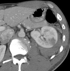
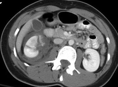
Grade III renal injury. Hypoechoic lesion in the left kidney after an MVA.

Grade V renal injury. Note the lacerations through and through the left kidney with surrounding hematoma.
Management
  • As mentioned, most (~98%) require only bed rest and observation in stable patients, even in grade IV and V injuries.
  • However, falling blood counts (Hb), persistent gross hematuria, hemodynamic instability and multiple transfusion requirements may indicate ongoing bleeding.
  • Arteriography and embolization to be considered in selective cases. 
  • Unstable patients may need explorative laparotomy.
  • Nephrectomy to be considered in extensive unsalvagable injuries, especially in hemodynamically unstable patients. 
  • Preservation of vessels attempted in cases of solitary kidneys or bilateral non-functioning kidneys.
Complications of Non-operative Management
  • Ongoing bleeding.
  • Urinary extravasation, may present as ileus, abdominal flank mass or discomfort. Urinomas, most resolve spontaneously.
  • Perinephric abscess.
  • Hydronephrosis
  • Arterio-venous fistula
  • Pseudoaneurysm
  • Pyelonephritis
  • Renal calculi
  • Hypertension, may be delayed.
Follow-up
  • Most have normal renal function and without hypertension.
  • The above complications need to be sought out.
  • Further imaging (ultrasonography to reduce radiation exposure) may be required if complications develops. 
Challenge:

Describe the images:


References:

Coran, Arnold G., et al., Pediatric Surgery, 7th Ed., Elsevier


Other future related topics to write on:
  • Management of trauma in children.
  • Ureter, bladder and urethral injuries.
  • Operative management.
  • Wilm's tumor.
  • Management Congenital anomalies.

Friday, January 4, 2013

Congenital Diaphragmatic Hernia: Short Notes

-->

Incidence

1 in 2000 - 5000 live births (unknown Malaysian figures)
Bochdalek hernia most common, left sided (80%)
Morgagni hernia (2%) usually in older children

Embryology

-       Diaphragm fusion involves 4 layers:
o   Septum transversum anteriorly
o   Pleuroperitoneal membrane dorsolaterally
o   Esophageal mesentry
o   Muscular portion from intercostal muscles
-       Starts at 4th week of gestation
-       Completes at 8th week of gestation
-       Lungs develope as a derivative from foregut and forms a diverticulum. Two lung buds form at around 4th week of gestation.
-     Airway differentiation developes up to 24 weeks of gestation. This is followed by appearance of pneumocyte I and II, the later responsible for secreting surfactant.

-     Vascular deveopement occur concurrently with lung growth. 

Pathology:
-       Gut herniates through the yolk sac and returns into the abdominal cavity at 8 – 9th week of gestation.
-       Failure of closire of diaphragm results in hernia
-       May cause obstruction to the esophagus (resulting in polyhydroamnios)
-       Circulation maintained as fetus is dependant on utero-placental-fetal circulation
-       After birth, adult type of circulation unable to be achieved due to persistent raised pulmonary pressure and reduction in pulmonary blood flow.
-       This results in right to left shunt resulting in deoxygenated blood circulating in the system and results in increase in pulmonary pressure.

Diagnosis

Antenatal:
Ultrasound
-       Stomach or other abdominal organ in the fetal thorax
-       Polyhydromnios (80%)

At birth:
-       Respiratory distress
-       Scaphoid abdomen (need high index of suspicion)
-       Asymmetrical distended chest wall
-       Shifted heart sounds (‘Dextrocardia’ is CDH until proven otherwise)
-       Absent breath sounds
-       Hypotension and peripheral hypoperfusion (reduced venous return from caval compression)
-       Chest X-ray: Loops of intestine or stomach in chest wall. Clearer if NG tube inserted.
-       Contrast studies might be indicated in certain cases but usually not necessary.

Some late presentations present with chronic lung disease, recurrent coughs, pneumonia.

Notice the heart on the right side and the nasogastric tube tip ending in the thorax.
 
Associated Anomalies
High incidence of lethal congenital anomalies asociated with CDH

-       Skeletal defects (32%)
-       Cardiac anomalies (24%)
-       Tracheobronchial anomalies (18%)
-       Neural tube defects

Differential Diagnosis:
Eventration of the diaphragm (weakness of the diaphragm ie. Phrenic nerve injury)

Management
Antenatal:
-       Tertiary center delivery with well equiped NICU and pediatric surgery support.
-       Transport to be made in-utero.
-       Parent counselling
-       Amnicentesis for karyotyping if suspected congenital anomaly
-       Delivery as indicated, not necessary for Caesarian section
-       In-utero intervention remains experimental

Delivery:
-       NBM
-       Ryles tube insertion
-       Prompt intubation, avoid bagging as may distend stomach and worsen condition. Difficulty to ventilate in unknown cases should have high index of suspicion.
-       Avoid excessive bagging as high risk of barotrauma and pneumothorax.
-       Adequate ventilation, pulmonary hypertension and pulmonary hypoplasia is the main complication of CDH and requires aggressive ventilatory support.
-       Adequate fluid resuscitation as hypotension may develop due to pulmonary hypertension.
-       Prompt surgical intervention not needed. Stabilization of patient and delayed surgery is preferred but lacking of controlled studies.
-       Extracorporeal membrane oxygenation (ECMO) in certain selected cases.


Surgery:
-       Approach: Subcostal or thoracotomy
-       Reduction of herniated organs
-       Primary repair
-       Placement of prosthesis over repaired diaphragm
-       Muscular and fascial flaps may be used to close repaired diaphragmatic defects.
-       Closure of abdominal incision might need to be done in two phases as there might not be enough ‘space’ for the returned abdominal organs. Silo might be necessary while awaiting for abdominal wall to expand for closure later.
-       ECMO support if necessary during surgery, but high risk of mortality from bleeding.
-       Chest tube not routine and only if indicated (pneumothorax)


Friday, December 28, 2012

Hydrops Fetalis: Workup

Historically, hydrops were first associated with fetal anemia caused by destruction of fetal red blood cells by antibodies produced by Rh negative mothers in response to antigenic exposure of a Rh positive fetus, usually from the first pregnancy. What result is a severely edematous fetus which characterizes this condition. In today's era where blood group is screened and Rh alloimmunization is prevented with anti Rh(D) immunoglobulin, the majority of causes of hydrops are non-immune related (~90%).

There is a whole list to the causes of hydrops. Typing every single condition would not be beneficial. I feel that work-up to the cause of hydrops should be aimed to detect maternal conditions that may lead to hydrops as well as anomalies that may recur. This is to prevent recurrence, if possible, in future pregnancies as mortality of neonates with hydrops is at 50% depending on the cause of hydrops. 

DIAGNOSIS

Diagnosis are usually made antenatally with detection of the following:
  • Skin edema
  • Scalp edema
  • Ascites
  • Pleural effusion
  • Pericardial effusion (most difficult to see)
CAUSES

I will not list all the possibilities but most of the common and important ones:

Immune-mediated:
Rhesus alloimmunization

Nonimmune causes:

Congenital anomalies
- Cystic hygroma
- Turner's (XO monosomy)
- Trisomies (21 most common)

Hematological
- Alpha thalassaemia
- Massive fetomaternal hemorrhage

Cardiac anomalies (all sorts)

Thoracic anomalies
- Congenital cystadenomatoid malformation (CCAM)
- Any mass/lesions that may cause increase thoracic pressure

Infections
- TORCHES (Toxoplasmosis, rubella, cytomegalovirus, Herpes simplex, syphilis [most common])
- Parvovirus B19

Metabolic Disorders
ie. Gaucher's disease, Tay-Sach's disease, sialidosis, generalized (GM1) gangliosidosis

Twinning
- Twin-twin transfusion syndrome

Others
- Skeletal dysplasias


WORK-UP

Antenatal

As usual, history from the mother should be obtained when hydrops is first detected. Family history of genetic anomalies, consanguineous marriage should be obtained. Recurrence are more likely in families with such histories ie. Alpha thalassaemia, metabolic disorders (which most are autosomal recessive), congenital anomalies.

History of exposure to infections may also be beneficial.

Rhesus negative mothers should always be ruled out.

Other associated anomalies may be looked for and detected during the initial ultrasound or repeated in more expert hands later. Amniocentesis for fetal karyotyping may be obtained after counselling. If infection is suspected, amniotic fluid may be sent for cultures or PCR if facilities are available. 

Postnatal

Evaluation of the neonate post-delivery should always be after initial resuscitation as these fetus tends to have turbulent births.

Relevant physical examination:
  • Cyanosis - may indicate underlying structural heart anomaly
  • Hepatosplenomegaly - metabolic disease, congenital infections
  • Features of Turner's, Down's and other features of chromosomal anomalies
  • Congenital anomalies - cystic hygroma, dysplasia
  • Hypotonia - metabolic disorders, dystrophies
  • Placenta - in case of TTTS
Investigations:
  • FBC - To rule out anemia or determine severity of anemia
  • PBF - To look for hemolysis
  • Kleihauer-Betke test - if fetomaternal hemorrhage suspected
  • ECG - to rule out arrythmias
  • Echocardiography - to rule out cardiac anomalies
  • Chest x-ray (Pleural effusion, CCAM)
  • Infection screen (TORCHES)
  • Karyotyping (if not yet done antenatally and if suspicious of chromosomal anomalies)
  • Metabolic screen (for metabolic diseases)
Management are then commenced according to cause and prognosis.

Postmortem:

If the fetus delivers as a stillbirth or had an early neonatal death, postmortem may be important to determine underlying anomalies or condition.


Determining the cause of hydrops is important to rule out causes that are potentially preventable or predictable, especially those associated with chromosomal anomalies. Future pregnancies may be followed-up more carefully with anticipation of treatment better. Mothers should always be counselled for possibilities for recurrence and next pregnancy planned well.

As mentioned this article only covers some of the causes and work-up for the hydrops patient. Mode of delivery, management of the fetus is not discussed in details here but both depends on the cause of hydrops and after further discussions with the parents.


References:
Creasy, Robert K., et al., Maternal Fetal Medicine, Principles and Practice, 6th Ed.
Speer, Micheal E., Postnatal Care of Hydrops Fetalis, Up-to-Date Article, 2012.

Sunday, December 23, 2012

Neonatal Resuscitation: The Basics

Fortunately most term babies are born without complications or needing help for resuscitation. About 10% do and that's a significant number. Basic knowledge of neonatal resuscitation must be available to all medical staffs (doctors and nurses) working in the labour room or the emergency department for that matter as mothers in advanced labour may just pop-up in the emergency department in the middle of the night.

The algorithm above pretty much sums up the steps of neonatal resuscitation covering even babies whom are delivered 'normal' without complications. There are many other variations but this is the one that I'm most used to.

Most times, the outcome of delivery can be roughly predicted even before birth itself. Knowledge of the patient's history helps a lot in anticipating outcome of delivery.

The initial step as in the box above emphasize the recognition of a baby in need of resuscitation. Four basic questions must be answered:
  • Is the baby term?
  • Is the liqour clear?
  • Is the baby crying or breathing?
  • Is the muscle tone good?
If any of the above is NO, then the next step of resuscitation is required. 

Even the first two questions above can be answered before delivery and can be anticipated. With anticipation comes preparation and with good preparation hopefully better outcome may be borne out of the delivery.

If all those answered above are YES, then the baby maybe passed on to the mother unless otherwise contraindicated, for skin to skin contact and initiation of breast feeding and routine care.

Another interesting note is that delayed cord-clamping should be practiced (as late as 1 minute after delivery) as there is evidence of reduced incidence of transfusion and increased iron storage in babies with delayed cord-clamping.

INITIAL STEPS TO RESUSCITATION

Below are the basic initial steps for resuscitation. All these steps are to be done ideally within 30 minutes and the baby reevaluated afterwards to assess success of resuscitation and need to proceed with the next step.

Provide warmth and Dry
- Baby is to be placed under radiant warmers.
- If baby is preterm, best to be wrapped in plastic wrappings as preterms are more likely to lose heat more easily

Position and maintain airway
- Positioned in sniffing position.

Stimulate
- Only back stimulation is necessary up to 2 to 3 times.
- Slapping the feet of babies should not be in practice today.

*Clear airway
- Latest guidelines from WHO states that babies whom are breathing on their own should not have regular suctioning, even if meconium was present.
- Suctioning should only be done if there are evidence of excessive secretions.
- There is no evidence in improved outcomes in babies suctioned intrapartum (after delivery of head, before delivery of shoulder) when meconium is present.
- If meconium is present and baby is not *vigorous, direct-suctioning with the meconium aspirator should be initiated straight away, even before positive pressure ventilation is performed.
- Direct suctioning is done until airway produces clear returns or baby becomes bradycardic.

*vigorous = not breathing, limp, heat rate <60 bpm.


POSITIVE PRESSURE VENTILATION (PPV)

- If the baby is still apneic or heart rate is less than 60 bpm after the initial steps, positive pressure ventilation should be initiated.
- 40 to 60 cycles of breath should be given per minute.
- Ensure there is chest rise. If not, reposition in sniffing position.
- Intubation is to be considered if unable to ventilate.
- Excessive pressure or 'bagging' must be avoided for risk of pneumothorax.
- Heart rate is to be evaluated after 60 seconds of PPV with target > 100 bpm.
- SpO2 monitoring may be attached during the resuscitation, best SpO2 probe to be placed at pre-ductal areas ie. right hand. One must be aware that oxygen saturation is not maximal in the newborn till at least 10 minutes after delivery.

Preductal SpO2 According to Duration after Birth
  • 1 minute - 60-65%
  • 2 minutes - 65-70%
  • 3 minutes - 70-75%
  • 4 minutes - 75-80%
  • 5 minutes - 80-85%
  • 10 minutes - 85-95%

CHEST COMPRESSIONS

- PPV and adequate ventilation should be done before initiation of chest compressions.
- Chest compressions done if heart rate < 60 bpm.
- Best to be done with 2 thumbs on the sternum with the other fingers around the baby for better control of pressure.
- Depth of compression should be 1/3rd of the AP diameter of the baby.
- Ratio of breath to compressions should be 1:3.
- 2 finger technique done if space is needed for procedures ie. insertion of umbilical catheters.
- Intubation must be considered at this stage if not earlier.

MEDICATIONS

- Rarely are medications need in neonatal resuscitation (as compared to adult resuscitation),
- If compressions are ineffective, IV adrenaline 0.01 - 0.03 mg/kg is to be given via intraumbilical venous catheter. If not, adrenaline 0.05 - 0.1 mg/kg may be administered through an endotracheal tube.
- Adrenaline should be diluted in 1:10000 (0.1 mg/mL)
- Adrenaline may be repeated every 3 to 5 minutes if heart rate remains < 60 bpm.
- IV bolus of N/S or HM 10 mL/kg may be given in suspected hypovolemia or anemic babies.
- Naloxone should not be given routinely.


Other Points: 
- Remember that resuscitation of the neonate should NOT be done alone.
- Always call for help of someone more experienced.
- Know what is available ie. laryngeal mask airway may be beneficial
- Know when to quit (Perhaps I'll write about this in other articles)
- And always inform the mother or parents of what was done and why and the outcome.
- Remember documentation.


That's it for now. This is just a 'short-note' of mine that I hope will be beneficial for me and to whoever that reads this. It's definitely incomplete but I hope will cover more of the basic stuff. Future related articles that I'll be posting may be on intubations, UVC insertions, other methods of ventilations, and withdrawing treatment, all important aspects of neonatal resuscitation. Let's hope that I've obtained at least a certificate for NRP by then :D


References:
- Neonatal resuscitation Textbook 5th Edition.
- Special Report - Neonatal Resuscitation, 2010 American Heart Association Guidelines for Cardiopulmonary Resuscitation and Emergency Cardiovascular Care, PEDIATRICS, Journal of American Academy of Pediatrics
- WHO Guidelines on Basic Newborn Resuscitation 2012.

Friday, December 21, 2012

Pilot

My birding blog Les'oiseoux seems to be the most active. Because birding is a passion. If not yet obsession :p Why not with my career? I have been reading but not really applying and reproducing. Why not why not?

It's been about 5 months into my MOship in O&G in Sabah's Women and Children Hospital. I have to say I have come some ways and come to realize that I like obstetrics, especially the fetal aspect of it. If only O&G was without the G...

My hope is that this blog covers on inputs and articles on children. Be it on diseases, activities, or hopefully later my own kids.

May Allah bless my efforts and those of others.