Showing posts with label short notes. Show all posts

Tuesday, July 9, 2013

Neonatal Stridor: Short Notes


Neonatal Stridor

Stridor is a harsh, raspy breath sounds heard during breathing that may indicate an upper airway obstruction. It may occur during inspiration, expiration or may be biphasic depending on the location of the obstruction.

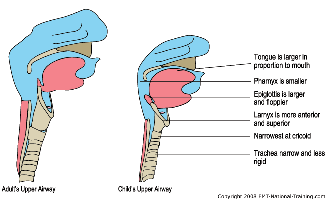
Anatomic and Physiologic Considerations

-       Smaller caliber of airway
-       Higher position and longer epiglottis
-       Lower total lung capacity
-       Increased functional residual capacity dependant on:
o   Increased respiratory rate
o   Active glottic narrowing during expiration
o   Use of expiratory muslces for respiration


Assessment

Babies with stridor should be assessed further for any evidence of respiratory distress:

-       Cyanosis
-       Chest retraction
-       Tachypnea
-       Grunting
-       Nasal flaring

Some may present with difficulty in feeding, recurrent choking or cough.

Investigations:

-       Flexible direct laryngoscopy
-       Rigid direct laryngoscopy

Differential Diagnosis

Supraglottic

-       Laryngomalacia (Commonest cause)


Omega shaped epiglottis


Glottic

-       Vocal cord paralysis (Second commonest cause)
-       Laryngeal web

Subglottic

-       Subglottic stenosis
-       Subglottic hemangioma
-       Tracheomalacia
-       Trachea stenosis with complete tracheal ring
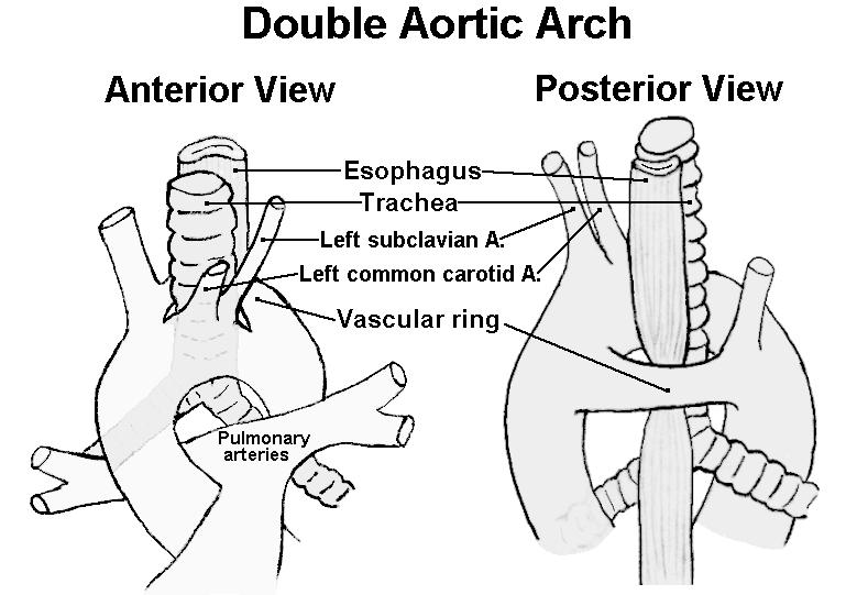
-       Pulmonary vascular ring


 Some websites for demonstrations and information:


Tuesday, April 30, 2013

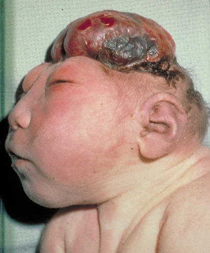
Anencephaly: Short Notes


Definition:
Absence of the calvaria above the bony orbits. May be associated with absence of cerebral hemisphere.

Incidence:
1/1000 live births.

Pathogenesis:
Failure of closure of cephalad neural tube at 25 - 27 days post conception.

Risk of Recurrence:
2 - 4 % with 1 affected siblings. 10% with 2 affected siblings.

Diagnosis (Antenatal):
  • Absent bony calvaria
  • Orbits well visualized
  • Absence of supratentorial brain
  • Residual brain
  • Associated polyhydromnios, due to decreased fetal swallowing
  • Associated spina bifida
Associated Anomalies:
  • Spina bifida
  • Urinary tract anomalies
  • Cleft lip / palate
  • GIT anomalies (ie. omphalocoele, intestinal aganglionosis)
  • Cardiac anomalies

Prognosis:
Incompatible with life. Some may live up to a week postnatally.

Prevention:
Folic acid supplements, recommended 4 mg per day a month preconception and 3 months after.
With-holding anticonvulsants (folic acid antagonist, ie. valproate, carbamezapine, phenytoin, trimetoprim)


References
1. Nyberg, David A., et al., Diagnostic Imaging of Fetal Anomalies, Lippincott Williams & Wilkins, 2003.

2. Tomita T., et al., Anencephaly, UpToDate Article, 2012.

Future Topics:
- Folic acid in prevention of neural tube defects.
- Feasibility of organ donation of anencephalic babies
- Spina bifida and other neural tube defects.


Monday, March 25, 2013

Sacroccocygeal Teratoma: Short Notes


Dr Vijay called me up to show a ultrasound picture of a patient he was seeing. The patient was referred for twins. What was revealed was instead a sacrococcygeal mass. A SCT, sacrococcygeal teratoma.


Born 18 May 2013, LSCS at 34 weeks.

************************************************************

SCT is a spectrum of germ cell tumors most commonly seen in the pediatric age groups.

Most germ cell tumors manifest extragonadally (sacrococcygeal, mediastinal, retroperitoneum) compared to adults where germ cell tumors present at gonadal areas (ovary, testes).

Extragonadal germ cell tumors are believed to arrive from arrested or abnormal migration of primordial germ cells to the genital ridge.

SCT are mostly benign.

Female predominance 4:1

Classification (Altman's Study)


Type I - Predominantly external
Type II - external with intrapelvic extension
Type III - Visible externally but predominantly internal
Type IV- Entirely presacral

- Type I and II constitutes about 80% of cases.
- Type III and IV (hidden types) have increased rates of malignancy.


Diagnosis and Manifestations

Prenatally

- Detected via ultrasound scans.
- May present as hydrops fetalis with polyhydromnios due to fetal blood shunting to tumor due to arteriovenous malformations in the tumor.

Perinatally

- Sacrococcygeal tumor

Older Children

- Sacroccocygeal tumor
- Urinary or bowel compression symptoms


-Alpha fetoproteins are elevated but should return to normal by 9 months of age.

Currarino Triad:
- Presacral teratoma
- Anal stenosis
- Sacral defect

Prognosis

- Reported 77% survival in antenatally diagnosed SCT.
- Only 14% survival for those with hydrops.
- A study reported 100% survival in lesions less than 10 cm, 48% in more than 10 cm.


Management:

Pre-op assessment:
MRI, CT scan or ultrasonography.

Resection
- Coccyx to be removed as recurrence rate is at 37% if not removed.

If deemed unresectable:
- Biopsy
- Neoadjuvant chemotherapy (cisplatin, bleomycin)

Adzick et al. was able to resect the tumore in-utero in a patient manifesting with polyhydromnios and hydrops.

Future Topics:

- In-utero surgery
- Surgical and post-op management
- Other germ cell tumors


References:

1. Coran, Arnold G., et al., Pediatric Surgery, 7th Ed., Elsevier. 

2. Nyberg, David A., et al., Diagnostic Imaging of Fetal Anomalies, Lippincott Williams & Wilkins, 2003.

Sunday, March 10, 2013

Hirschsprung Disease: Short Notes

Disease caused by absence of ganglions in the myenteric (Auerbach) and submucosal (Meissner) plexus causing functional intestinal obstruction.

Long-segment Hirschsprung: Aganglionic bowel proximal to midtransverse colon 

Embryology and Etiology

Ganglion cells originate from the neural crest tissues by 13 weeks postconception from proximal to distal GIT.

2 theories:
  • Failure of migration of neural crest tissues.
  • Failure of migrated ganglionic cells to mature and proliferate
 Mutation of RET proto-oncogenes

Associated Syndromes

Down syndrome
Neurocristopathy syndromes (ie. Waardenberg-Shah syndrome)
Malrotation
Congenital heart disease
Urinary tract anomalies
Central nervous system anomalies
Congenital central hypoventilation syndrome (Ondine curse)

Clinical Features

Neonates:
  • Delayed passage of meconium after 24 hours
  • Abdominal distension
  • Bilious vomiting
  • Feed intolerance
- Chronic constipation
- Diarrhea, fever, abdominal distension (Hirschprung-associated enterocolitis [HAED])


Investigations

Radiographic
- Abdominal X-ray
- Barium enema

  • Transition zone between normal and aganglionic bowel
  • Reversed recto-sigmoid index (< 1.0)
Anorectal Manometry
  • Recto-anal inhibitory reflex (RAIR). Reflex that causes the internal anal spinchter to relax when the rectum is distended.
  • Balloon inserted into the rectum and inflated. If reflex present detected by sensor.
  • Test more useful with older children.
Rectal biopsy
  • Absence of ganglionic cell
  • Hypertrophied nerve trunk
  • Acetylcholinesterase staining
  • Absence of calcitonin on immunochemical testing with calretinin
Management

Resuscitation
  • Fluids
  • Nasogastric tube
  • Antibiotics
Operative

May be done through laparotomy or more popular, pull-through surgery via the anus.
  • Swenson (end-to-end anastamosis of the ganglionic segment with the internal anal sphincter. Rectum is excised)
  • Soave (As Swenson but rectum is preserved becoming the outer 'cuff' of the pulled ganglinic segment)
  • Duhamel (The ganglinic segment is joined to the rectum in a end-to-side anastamosis)
Colostomy to consider:
  • Enterocolitis
  • Perforated bowel
  • Very dilated proximal bowel
Surgery for Long-segment Hirschsprung:
  • Ileostomy
  • Small bowel pull-through to anastamose with the anal sphincter
  • Side-to-side anastamosis between small bowel and large bowel (to preserve absorptive roles). Several techniques: Martin and Kimura
 Post-op Care
  • Anal calibration 4 to 6 weeks
  • Buttock care with barrier cream to prevent perineal wound breakdown

  
 References:

1. Coran, Arnold G., et al., Pediatric Surgery, 7th Ed., Elsevier

Both pictures from http://radiopaedia.org/cases/hirschsprung-disease