Saturday, May 11, 2013

Autism: DSM IV Criteria

A total of 6 (or more) from (1) , (2) , (3), with at least 2 from (1), and 1 from each (2) and (3).

1. Qualitative impairment in social interaction, as manifested by at least 2 of the following:

a. Marked impairment in use of multiple nonverbal behaviors, such as eye to eye gaze, facial expression, body postures, and gestures to regulate social interaction.
b. Marked failure to develop peer relationships appropriate to developmental level.
c. Lack of spontaneous seeking to share enjoyment, interests, or achievements with other people (ie. lack or showing, bringing, pointing object of interest).
d. Lack of social or emotional reciprocity.

2. Qualitative impairments in communication, as manifested by at least 1 of the following:

a. Delay in or total lack of development of spoken language (not accompanied by an attempt to compensate through alternative modes of communications such as gesture or mimes).
b. In individual with adequate speech, marked impairment in ability to initiate or sustain conversation with others.
c. Stereotyped and repetitive use of language or idiosyncratic language.
d. Lack of varied, spontaneous make-believe play or social imitative play appropriate to developmental level.

3. Restricted, repetitive, and stereotyped patterns of behavior, interest and activities as manifested by at least 1 of the following:

a. Encompassing preoccupation with 1 or more stereotyped and restricted pattern of interest that is abnormal in either intensity or focus.
b. Apparently inflexible adherence to specific, nonfunctional routines or rituals.
c. Stereotyped and repetitive motor mannerism (ie. hand or finger flapping or twisting or complex body movements)
c. Persistent occupation with parts of objects.

B. Delay or abnormal functioning in at least 1 of the following areas with onset < 3 age year: (1) social interaction, (2) language as used in social communication, or (3) symbolic or imaginative play

C. The disturbance is not better accounted by Rett disorder or childhood degenerative disorder.


***********************************

This article may be a bit out-dates when DSM V comes out with new changes. Asperger's and Persistent Developmental Disorder - Not Otherwise Specified (PDD-NOS) will be grouped together with Autism under Autistic Spectrum Disorders with various levels.

Asperger's differ only in terms of language skills which is mostly intact.

Autism has no association with MMR vaccination.

Reference:

1. Kliegman, Robert M., et al., Nelson Textbook of Pediatrics, 18th ed., Elsevier Saunders.

Future Topics:
- Management
- DSM V!
 

Tuesday, April 30, 2013

Anencephaly: Short Notes


Definition:
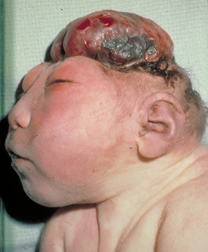
Absence of the calvaria above the bony orbits. May be associated with absence of cerebral hemisphere.

Incidence:
1/1000 live births.

Pathogenesis:
Failure of closure of cephalad neural tube at 25 - 27 days post conception.

Risk of Recurrence:
2 - 4 % with 1 affected siblings. 10% with 2 affected siblings.

Diagnosis (Antenatal):
  • Absent bony calvaria
  • Orbits well visualized
  • Absence of supratentorial brain
  • Residual brain
  • Associated polyhydromnios, due to decreased fetal swallowing
  • Associated spina bifida
Associated Anomalies:
  • Spina bifida
  • Urinary tract anomalies
  • Cleft lip / palate
  • GIT anomalies (ie. omphalocoele, intestinal aganglionosis)
  • Cardiac anomalies

Prognosis:
Incompatible with life. Some may live up to a week postnatally.

Prevention:
Folic acid supplements, recommended 4 mg per day a month preconception and 3 months after.
With-holding anticonvulsants (folic acid antagonist, ie. valproate, carbamezapine, phenytoin, trimetoprim)


References
1. Nyberg, David A., et al., Diagnostic Imaging of Fetal Anomalies, Lippincott Williams & Wilkins, 2003.

2. Tomita T., et al., Anencephaly, UpToDate Article, 2012.

Future Topics:
- Folic acid in prevention of neural tube defects.
- Feasibility of organ donation of anencephalic babies
- Spina bifida and other neural tube defects.


Omphalocoele & Gastrochisis

Both are quite common abdominal wall defects afflicting neonates. Omphalocoele and gastrochisis were once thought to be the same entity until quite recently. Gastrochisis was once thought to be a ruptured omphalocoele. Now it is well known that this is not the case.

Other disease of abdominal wall defect includes cloacal exstrophy (epispadias), pentology of Cantrell and cordis extrophy but will not be covered in this article.

Definitions

Omphalocoele
This is a large defect in the anterior abdominal wall when amniotic membrane covers protruding midgut components which may include the liver, gonads and spleen.


Gastrochisis
This is usually a smaller defect with protrusion of midgut components without membrane covering. The defect is usually to the right of the umbilicus.

In both defects, the rectus abdominis muscles are intact.


Umbilical hernia differs from omphalocoele in the it is covered by skin instead of amniotic membrane. It also usually appears not at birth but much later after.


Prevelance

Gastrochisis is more common than omphalocoele. Incidence is about 2 - 4.9 per 10000 live births.

Omphalocoele has an incidence of about 1 - 2.5 per 10000 live births.

Embryology

The body cavities are enclosed by folding of the embronic disc, anteriorly, caudally, and right and left laterally. This will leave the yolk sac in the middle. This occurs at 3 weeks of gestation.

At 5 weeks, the gut 'herniate' into the yolk sac and develop within.

At 10 weeks, the gut retracts back into the peritoneal cavity.

At least this is whats supposed to happen. When there's a failure of the lateral fold to complete, omphalocoele develops with failure of retraction of the midgut.

Gastrochisis supposedly occur due to failure of developement of the yolk sac. The gut has no space to expands and ruptures out of the abdominal wall. This usually occur on the right side of the umbilicus, probably due to weakness at that side due to involution of the right umbilical vein at 4 weeks of gestation.

Associated Anomalies

Omphalocoele
  • Chromosomal ~20%
  1. Trisomy 18, 13, Triploidy, 45 X (Turner's)
  2. Aneuploidy
  3. Beckwith-Wiedemann syndrome
  • Cardiac ~ 7 - 50%
  • Central nervous system ~ 4 - 30%
  • Musculoskeletal ~ 4 - 25%
  • Genitourinary ~ 6 - 20%
  • Gastrointestinal ~ 3 - 20% (Less compared to gastrochisis, probably due to protective effect of covering sac.
Gastrochisis

More associated with midgut anomalies
  • Intestinal atresia ~ 7 - 30% (due to mesentric ischemia, likely due to compression by small abdominal defect)
  • GERD ~ 16%
  • Undescended testis ~ 15%

Antenatal Diagnosis

Sensitivity for detection with ultrasonography: 75% for omphalocoele, 83% for gastrochisis.

Omphalocoele
Centrally located, at base of umbilical cord insertion.
Presence of membrane.
Presence of liver (extracorporeal liver) carries poorer prognosis.
Associated with polyhydromnios (poorer prognosis)

Gastrochisis
Abdominal wall defect noted at right of umbilicus.
Variable amount of bowel protrusion.
Bowels usually thickened and edematous.
Assoicated with oligohydromnios
Associated with IUGR

Other anomalies should be sought out, especially for omphalocoeles.

Both have elevated serum and amniotic aFP and amniotic fluid acetylcholine (AChE) but more in gastrochisis (~80 more).

Amniocentesis may also help to rule out chromosomal abnormalities that may predict prognosis.

All above information combined may provide useful information to convey to parents and provide counseling as well as for discussion of prognosis.


Mode of Delivery

Controversial. No good RCT studies.

For gastrochisis, some reports less GIT complications with elective Caesarian section. Caesarian section recommended for large omphalocoele to prevent rupture of sac.

No benefit in earlier delivery unless if obstetrically indicated. Preterm delivery is associated with more complications associated with prematurity that may make management of the infant more difficult postnatally.

Termination of pregnancy should be considered and discussed with chromosomal abnormalities.

Delivery should ideally be done in a center with pediatric surgery expertise. Conditions of the bowels post-delivery is related to duration of time it takes between delivery and repair.

Management

Initial Care
  • Keep NBM
  • Keep warm
  • Intestinal decompression with nasogastric tube and meconium evacuation
  • Adequate hydration
  • Prevent heat loss (especially important in gastrochisis where the bowels without sac are exposed, wrap with plastic or kitchen wrap).
  • Respiratory support, ventilator readily available.
  • Other anomalies excluded, especially cardiac lesions with omphalocoele.
Surgical Care


Omphalocoele
  • The sac is cleaned with antiseptic.
  • Attempt be made at manual reduction. May not succeed wholly at first. Incision around the defect is made and then the sac excised.
  • Abdominal wall may be stretched posterior-to-anterior and bowel be replaced first followed by the liver to wedge the bowel in.
  • The anterior abdominal wall is closed with sutures applied on all layers of the rectus including the muscle. After the closure is done without tying off the knot, an attempt is made to ventilate the patient. Closure is safe if patient is able to be ventilated with peak inspiratory pressure of less than cm H2O. 
  • If unable to reduce without closing the abdominal wall too tightly, a prosthetic silo is applied and the bowel reduced weekly.
  • Any atresia or other bowel anomalies found may be operated 6 weeks after initial surgery.
Gastrochisis
  • Similar principle to omphalocoele.
  • Bowels are reduced and primary closure.
  • While waiting, the bowel may be kept in a prosthetic silo. Some centers attempt to reduce bowel bedside.
Post-operative Care
  • Continued ventilation.
  • Continued hydration with urine output monitoring.
  • Antibiotics.
  • Bowel function may be delayed for up to 3 weeks. Any longer a contrast study may be indicated.
  • Monitoring of complications related to respiratory difficulties. Sutures may need to be released and gradual reduction be made.
Complications

Short Term

  • Associated anomalies (especially in omphalocoele).
  • Associated with prematurity (especially in gastrochisis; hypoglycemia, respiratory distress syndrome, electrolyte imbalance, heat loss).
  • Associated with tight closure causing respiratory distress.
Long Term

Small bowel obstruction.
*Absence of umbilicus (social distress).
Poor weight gain
Low IQ

Most of these complications vary in frequency depending on where the study is conducted.

References:

1. Coran, Arnold G., et al., Pediatric Surgery, 7th Ed., Elsevier. 

2. Hutson, John M., et al., Jones' Clinical Pediatric Surgery Diagnosis and Management, 6th ed., Blackwell Publishing (2008).

3. Nyberg, David A., et al., Diagnostic Imaging of Fetal Anomalies, Lippincott Williams & Wilkins, 2003.

Future Topics:

Surgical management
Bladder extrophy
Pentalogy of Cantrell
Cordis ectopia

Tuesday, March 26, 2013

Salter-Harris Classification of Fracture: Revision


This classification is for fractures in pediatric age group that involves the physeal growth plate.

Histology

The germinal layer of the growing bone is on the epiphysis. Cells proliferate towards the metaphysis with more mature cells at that end ossifying.

Damage to the growing or proliferating part of the growth plate may result in premature halt of cell growth and bone growth.


Classification (Original)

Type I - fracture through the hypertrophic part of the physeal plate without involving the metaphysis or epiphysis.

Type II - fracture involving the metaphysis and physeal plate.

Type III - fracture involving the epiphysis and physeal plate.

Type IV - through and through fracture involving both epiphysis and metaphysis through the growth plate.

Type V - Crush injury of the physeal plate.

Other rarer types:

Type VI - Injury to perichondral stuctures.

Type VII - Isolated injury to epiphyseal plate.

Type VIII - Isolated injury to metaphysis with possible injury to endochondral ossification.

Type IX - Injury to periosteum that may interfere with membranous growth.

Prognosis and Complications

Type I and II are associated with better prognosis and usually require only conservative management.

Type III and above may require surgical intervention because of the potential complications of limb length discrepanciesor shortening, angulation of limbs, and intraarticular involvement that may result in chronic disability.

Future Topics:

- Fracture management in pediatrics
- Fractures specific to pediatric age groups (ie. Supracondylar fracture)
- CRITOE: Ossification in children

Credits:

Bevan, Chad, Slater-Harris Fractures, Power Point Slide, 2007.

Monday, March 25, 2013

Sacroccocygeal Teratoma: Short Notes


Dr Vijay called me up to show a ultrasound picture of a patient he was seeing. The patient was referred for twins. What was revealed was instead a sacrococcygeal mass. A SCT, sacrococcygeal teratoma.


Born 18 May 2013, LSCS at 34 weeks.

************************************************************

SCT is a spectrum of germ cell tumors most commonly seen in the pediatric age groups.

Most germ cell tumors manifest extragonadally (sacrococcygeal, mediastinal, retroperitoneum) compared to adults where germ cell tumors present at gonadal areas (ovary, testes).

Extragonadal germ cell tumors are believed to arrive from arrested or abnormal migration of primordial germ cells to the genital ridge.

SCT are mostly benign.

Female predominance 4:1

Classification (Altman's Study)


Type I - Predominantly external
Type II - external with intrapelvic extension
Type III - Visible externally but predominantly internal
Type IV- Entirely presacral

- Type I and II constitutes about 80% of cases.
- Type III and IV (hidden types) have increased rates of malignancy.


Diagnosis and Manifestations

Prenatally

- Detected via ultrasound scans.
- May present as hydrops fetalis with polyhydromnios due to fetal blood shunting to tumor due to arteriovenous malformations in the tumor.

Perinatally

- Sacrococcygeal tumor

Older Children

- Sacroccocygeal tumor
- Urinary or bowel compression symptoms


-Alpha fetoproteins are elevated but should return to normal by 9 months of age.

Currarino Triad:
- Presacral teratoma
- Anal stenosis
- Sacral defect

Prognosis

- Reported 77% survival in antenatally diagnosed SCT.
- Only 14% survival for those with hydrops.
- A study reported 100% survival in lesions less than 10 cm, 48% in more than 10 cm.


Management:

Pre-op assessment:
MRI, CT scan or ultrasonography.

Resection
- Coccyx to be removed as recurrence rate is at 37% if not removed.

If deemed unresectable:
- Biopsy
- Neoadjuvant chemotherapy (cisplatin, bleomycin)

Adzick et al. was able to resect the tumore in-utero in a patient manifesting with polyhydromnios and hydrops.

Future Topics:

- In-utero surgery
- Surgical and post-op management
- Other germ cell tumors


References:

1. Coran, Arnold G., et al., Pediatric Surgery, 7th Ed., Elsevier. 

2. Nyberg, David A., et al., Diagnostic Imaging of Fetal Anomalies, Lippincott Williams & Wilkins, 2003.

Thursday, March 14, 2013

Torsion of the Testes and It's Appendage: Review


Introduction

***TESTICULAR TORSION IS A SURGICAL EMERGENCY***

Many boys and men have lost their lives due to late diagnosis and treatment of this condition. It is not the most common cause of acute scrotum but is the most important as late management results in irreversible ischemia and gonadal necrosis. Lack of awareness and late presentation by patients and parents also contribute to delayed management of this condition.

Epidemiology

Testicular torsion may be divided to intratunical or intravaginal torsion and extratunical or extravaginal. This refers to the torsion occurring with the tunica vaginalis invaginating the testes or not.

Extratunical torsion occurs mostly during the perinatal period as the loose areolar tissue surrounding the testes predispose to torsion during descent. 70% occurs prenatally, while 30% occurs postnatally. Adherence of the testes to the surrounding tissues usually occurs around 6 weeks of age.

Intratunical torsion mostly occur with the puberty age group of boys (13 - 16 years). This might be related to testicular enlargement due to increased testesterone production during this period. The left testes is more frequently involved.

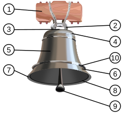
This mostly occur in the bell-clapper anomaly type of testes when the tunica vaginalis invaginate superiorly involving the spermatic cord, instead of just the epididymis and testes. In the normal testes, the posterior part of the testes is anchored to the scrotum preventing from much mobility. However when completely surrounded by the tunica vaginalis, the testes becomes more mobile at its axis through the spermatic cord. This anomaly usually occurs bilaterally requiring contralateral orchidopexy during surgery.

Bell-clapper: 5. Tunica vaginalis. 9. Testes. 

Angular relations between the testes and the tunica vaginalis. Testes lies more horizontal with higher invagination of the tunica.


Cryptochordism is also associated with higher incidence of torsion. Trauma and exercise causing higher cremesteric activity may also result in torsed testes.

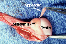
The commoner but less serious torsion of the testicular appendix (Hydatid of Morgagni) occurs also most often at puberty. The appendage is present in 90% of boys. In a review of 771 children with acute scrotum, 58% had torsion of the testicular appendix, 29% of the testes.


Rarely, the testes is torsed between it and the epididymis connected loosely.


Clinical Features

***Epididymo-orchitis is rare in children and adolescents***

The typical feature is sudden onset of pain at the scrotum or the ipsilateral iliac fossa. This may or may not be accompanied by nausea and vomiting. Beware that some may present with gradual onset of pain leading to delayed diagnosis. Resolving pain may indicate spontaneous resolution of the torsion or worse, a necrotic testes.

Examination reveals inflammed testes which is tender to touch. The testes is usually high riding as well with absence of the cremasteric reflex. However absence of reflex should not be the only indicator as there are case reports of presence of this reflex in a case of testicular torsion. The presence of the bell-clapper anomaly makes the diagnosis more likely and indicated for surgical exploration. Secondary reactive hydrocele may make testicular palpation more difficult.


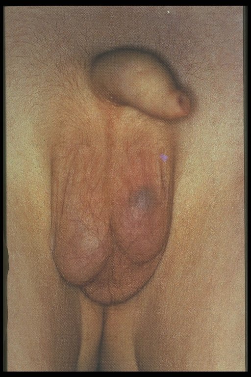
In a torsed testicular appendix, the pain is usually not as severe and more localized. A 'blue-dot sign' may be seen and point palpation is tender, while palpation of the whole testes is not. However, this is difficult to elicit once secondary hydrocele develop and the scrotum becomes edematous.


Blue-dot sign


Acute scrotum in an infant.


Investigations

***DO NOT LET DELAY IN OBTAINING IMAGING INVESTIGATION DELAY SURGICAL INTERVENTION AND MANAGEMENT***

Ultrasound doppler has a sensitivity of 88% and specificity of 90%. Absence of blood flow to the affected testes is diagnostic. Parenchymal echonegicity is also decreased although it may be increased after infarction. Echogenic, enlarged epididymis is an ancillary sign of testicular torsion. Ultrasound may also help differentiate from hydrocele, abscess, hematoma or tumor.

Torsed appendix usually presents as hyper-echogenic nodule between the epididymis and testes.

Epididymitis may present as hypervascular epididymis although torsion should not be excluded based on just this feature.

Radio-isotope scan may also help in accurately diagnosing testicular torsion. This is unfortunately not widely available.


Note the absence of blood flow in the left testes.


Management

***IF IN DOUBT OF DIAGNOSIS, SURGICALLY EXPLORE***

Manipulation of the testes has been attempted although most suggest that this is done only while waiting for preparation before surgical exploration. This is done in an 'open book' manner, rolling the testes laterally. Success of this maneuver usually relieves the pain spontaneously. This maneuver however is practically difficult to perform on a uncooperative child unless under anesthesia or adequate pain relief. Furthermore, if the torsion occurred in a lateral rotation, the maneuver may worsen the torsion.

Surgical exploration is warranted if diagnosis is in doubt or there is high suspicion of testicular torsion. Incision may be made paramedian, transverse or vertically through the scrotum. The torsed tested is untwisted.

Examination is done to see testicular viability. If testes is infarcted, orchidopexy is recommended in patients more than 10 years old, as there is risk of autoimmunization with spermatogonia as there is a breech between the blood-testes barrier which may affect fertility at a later age even when the contralateral testes is uninvloved. Some surgeons leave the necrosed testes in-situ in children less than 10 years old.

If the testes is viable, the tested is fixed (orchidopexy). The contralateral testes is also fixed as well, especially when the torsion is associated with the bell-clapper anomaly as this usually occurs bilaterally. It is recommended to use non-absorbable sutures in fixation of the testes tunica albugenia and dartos layer. Use of absorbable sutures are associated with recurrent torsion.
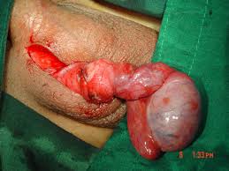

Torsion of the testes. The testes is still viable. Incision of the testes with presence of bleeding may confirm this.


In the torsed appendix of the testes, this is excised.

Necrosed torsioned testicular appendix.


Surgical management is more controversial in torsion in the neonates. 


Prognosis

Rate of testes salvage is inversely proportional with ischemia time. Testes is salvagable by 90% if exploration is performed within 4 - 6 hours after onset of symptoms, 50% if done at 12 hours, and 10% if present more than 24 hours.


Conclusion and Take-Homes

Testicular torsion is a surgical emergency which prompt exploration and treatment is important if the testes were to be salvaged. Diagnosis although may be made by imaging studies, should not cause delay in surgical exploration. The contralateral testes should be explored as well and orchidopexy performed with non-absorbable sutures.


References:

1. Coran, Arnold G., et al., Pediatric Surgery, 7th Ed., Elsevier 

2. Hutson, John M., et al., Jones' Clinical Pediatric Surgery Diagnosis and Management, 6th ed., Blackwell Publishing (2008).

3. Godbole, Prasad P., Testicular Problems in Children, Pediatric and Child Health, Vol 22:6, June 2012.

4. Govindarajan, Krishna K., Pediatric Testicular Torsion, Emedicine, Medscape article.

5. Various websites for the pictures in which none are mine.

Sunday, March 10, 2013

Hirschsprung Disease: Short Notes

Disease caused by absence of ganglions in the myenteric (Auerbach) and submucosal (Meissner) plexus causing functional intestinal obstruction.

Long-segment Hirschsprung: Aganglionic bowel proximal to midtransverse colon 

Embryology and Etiology

Ganglion cells originate from the neural crest tissues by 13 weeks postconception from proximal to distal GIT.

2 theories:
  • Failure of migration of neural crest tissues.
  • Failure of migrated ganglionic cells to mature and proliferate
 Mutation of RET proto-oncogenes

Associated Syndromes

Down syndrome
Neurocristopathy syndromes (ie. Waardenberg-Shah syndrome)
Malrotation
Congenital heart disease
Urinary tract anomalies
Central nervous system anomalies
Congenital central hypoventilation syndrome (Ondine curse)

Clinical Features

Neonates:
  • Delayed passage of meconium after 24 hours
  • Abdominal distension
  • Bilious vomiting
  • Feed intolerance
- Chronic constipation
- Diarrhea, fever, abdominal distension (Hirschprung-associated enterocolitis [HAED])


Investigations

Radiographic
- Abdominal X-ray
- Barium enema

  • Transition zone between normal and aganglionic bowel
  • Reversed recto-sigmoid index (< 1.0)
Anorectal Manometry
  • Recto-anal inhibitory reflex (RAIR). Reflex that causes the internal anal spinchter to relax when the rectum is distended.
  • Balloon inserted into the rectum and inflated. If reflex present detected by sensor.
  • Test more useful with older children.
Rectal biopsy
  • Absence of ganglionic cell
  • Hypertrophied nerve trunk
  • Acetylcholinesterase staining
  • Absence of calcitonin on immunochemical testing with calretinin
Management

Resuscitation
  • Fluids
  • Nasogastric tube
  • Antibiotics
Operative

May be done through laparotomy or more popular, pull-through surgery via the anus.
  • Swenson (end-to-end anastamosis of the ganglionic segment with the internal anal sphincter. Rectum is excised)
  • Soave (As Swenson but rectum is preserved becoming the outer 'cuff' of the pulled ganglinic segment)
  • Duhamel (The ganglinic segment is joined to the rectum in a end-to-side anastamosis)
Colostomy to consider:
  • Enterocolitis
  • Perforated bowel
  • Very dilated proximal bowel
Surgery for Long-segment Hirschsprung:
  • Ileostomy
  • Small bowel pull-through to anastamose with the anal sphincter
  • Side-to-side anastamosis between small bowel and large bowel (to preserve absorptive roles). Several techniques: Martin and Kimura
 Post-op Care
  • Anal calibration 4 to 6 weeks
  • Buttock care with barrier cream to prevent perineal wound breakdown

  
 References:

1. Coran, Arnold G., et al., Pediatric Surgery, 7th Ed., Elsevier

Both pictures from http://radiopaedia.org/cases/hirschsprung-disease