Epidemiology
- Injuries are classified into blunt and penetrating injuries.
- MVAs make up about 60% of the cause of renal injuries, followed by falls (20%) and sports injuries (10%).
- Sudden decelerations with flexion-extension movement in seat-belt is a known mechanism of injury
- In the USA, penetrating injuries are caused mostly by gunshot wounds (86%) followed by stab wounds.
- Associated injuries include liver, spleen and bowel injury. These injuries most be sought for and ruled out.
Certain features and differences in the child's kidneys make them more susceptible to injury compared to adults.
- Kidneys are relative larger compared to the size of the child's body.
- Position is much lower as well in the abdomen.
- Less protected due to decreased perirenal fat.
- Renal capsule and Gerota's fascia less well-developed.
- More mobile kidneys at the pedicle make it more susceptible to deceleration injuries.
- Retainment of lobulations increase risk of parenchymal distruption.
- Weaker abdominal wall muscles and poorly ossified rib cages make renal injury more likely.
- Congenital anomalies increases risk ie. hydronephrosis, horseshoe kidney, polycystic kidneys, renal tumors.
- Abdominal pain and flank pain.
- Flank echymosis
- Hematuria (absence does not exclude underlying injury)
- Associated injuries include lower ribs and lumbar vertabrae fracture.
- A lot of children are also asymptomatic
- FBC, Renal function
- Urinalysis (controversy in further radiological imaging if microscopic hematuria is present. Request for imaging must be correlated with the whole clinical picture).
- Contrasted abdominal CT-scan is standard for evaluation.
- Intravenous pyelography useful in unstable patient prior to op to determine functioning of 2 kidneys, extent of urinary extravasation, pedicle injuries.
- Ultrasound not sensitive in detecting parenchymal injuries. Useful for follow-ups to exclude urinomas, expanding hematomas, abscess, pseudoaneurysm.
 |
| From: Simple Medicine website, http://simple-med.blogspot.com/ |
American Association for the Surgery of Trauma (AAST) Grading for Renal Injury
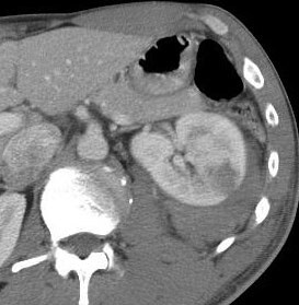
Grade I: Subcapsular, nonexpanding hematoma. Microscopic or gross hematuria with normal urologic studies in contusions.
Grade II: < 1 cm parenchymal depth of cortex laceration without urinary extravasation. Non-expanding hematoma confined to renal retroperitoneum.
Grade III: > 1 cm parenchymal depth of cortex laceration without urinary extravasation.
Grade IV: Laceration extending up to cortex, medulla and collecting system. Renal artery or vein injury with contained hemorrhage.
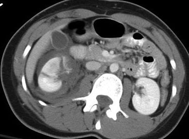
Grade V: Complete shattered kidney. Avulsion of renal hilum that devascularizes the kidney.
*Grading to standardize description for research and collection purposes.
**Generally the lower the grade I - III, the less likely need for operative intevention provided patient is stable.
***This grading is developed for adults that although may apply for children, but management should be based on case to case basis.
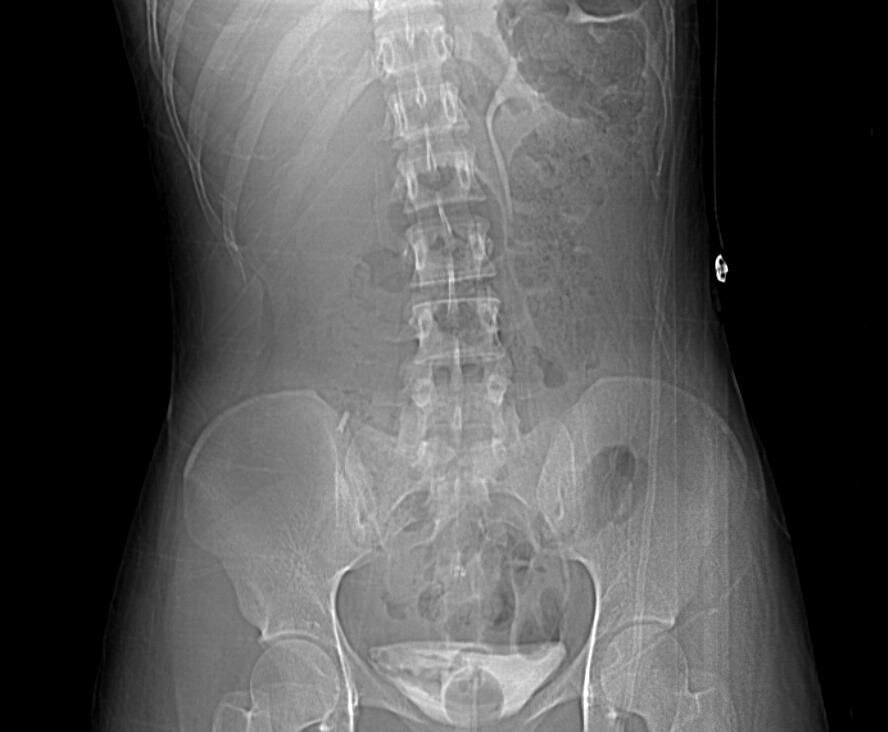
The following pictures are not only from pediatric cases.
 |
| Grade III renal injury. Hypoechoic lesion in the left kidney after an MVA. |
 |
| Grade V renal injury. Note the lacerations through and through the left kidney with surrounding hematoma. |
Management
- As mentioned, most (~98%) require only bed rest and observation in stable patients, even in grade IV and V injuries.
- However, falling blood counts (Hb), persistent gross hematuria, hemodynamic instability and multiple transfusion requirements may indicate ongoing bleeding.
- Arteriography and embolization to be considered in selective cases.
- Unstable patients may need explorative laparotomy.
- Nephrectomy to be considered in extensive unsalvagable injuries, especially in hemodynamically unstable patients.
- Preservation of vessels attempted in cases of solitary kidneys or bilateral non-functioning kidneys.
- Ongoing bleeding.
- Urinary extravasation, may present as ileus, abdominal flank mass or discomfort. Urinomas, most resolve spontaneously.
- Perinephric abscess.
- Hydronephrosis
- Arterio-venous fistula
- Pseudoaneurysm
- Pyelonephritis
- Renal calculi
- Hypertension, may be delayed.
Follow-up
- Most have normal renal function and without hypertension.
- The above complications need to be sought out.
- Further imaging (ultrasonography to reduce radiation exposure) may be required if complications develops.
Describe the images:


References:
Coran, Arnold G., et al., Pediatric Surgery, 7th Ed., Elsevier
Other future related topics to write on:
- Management of trauma in children.
- Ureter, bladder and urethral injuries.
- Operative management.
- Wilm's tumor.
- Management Congenital anomalies.

No comments:
Post a Comment