Monday, September 2, 2013

Obesity in Kids! Assessment

Childhood obesity has made headlines, most recently this month on Malaysian newspaper, 1 and was a dsitinction viva question in IIUM's latest professional exam as well. Malaysia is becoming a fatter nation with fat children. This is all linked to the standard of living and the way we live our lives.


As for kids becoming fat, most are not 100% their own fault. Parental styles contribute, family and cultural lifestyle, as well as peer pressure play roles in this 'disease' of fatness. Some (a lot) take offense to public comments on fatness as well.

Understanding the risks and complications may help in creating awareness in the general public. Only then can an effective counter-measure be implemented.


********************************************

Diagnosis and Assessment of Obesity

Diagnosis is based on the BMI centile charts in children and adolescence. This is in contrast to adults where the absolute value of BMI determine whether a person is obese.

Calculation of BMI is the same = Weight (kg) / [ Height (cm) ] square root.

Centile charts here.

Anything above 95% is considered OBESE.


What to do when you see a really fat kid in your clinic?

History Taking:

Family History:
- Fat parents and siblings
- Metabolic syndrome or cardiovascular disease in family

Antenatal and Postnatal History:
- GDM mother
- High birth weight
- Non-exclusive breastfeed
- Early introduction to sweetened fluids and food

Monster 5 kg.


Social History:
- Diet history: frequency of snacks, carbonated or sweet drinks, eating as a family, eating outside.
- Physical activity: frequency, types of activities, 'screen' time ie. TV, internet, playstation.
- Attitude towards obesity, and losing weight. 
- School and peer: Bullying, bully, taunting.

Symptoms (related to Complications or Comorbids, see later)


Physical Examination

Short statured (Cushing syndrome)
Acne (PCOS, Cushing syndrome) 
Acanthosis nigricans (Metabolic syndrome, PCOS)

Blood pressure

Abducent nerve palsy, papilloedema (Pseudoumor cerebri)

Striae (Cushing syndrome)
Abdominal pain (Gallstone)
Hepatomegaly (Non-alcoholic fatty liver disease)

Limited range of movement of hips (Slipped capital femoral epiphysis)
Bowing of legs (Blounts disease)


Investigations

Fasting lipid profile
Fasting sugar profile
Liver function test

Imaging as indicated according to complications.
Hip and lower limb x-ray
Abdominal ultrasound


Complications and Comorbids
 
 Respiratory System
  • Asthma
  • Obstructive sleep apnea
 Cardiovascular System
  • Hypertension
Endocrine System
  • Metabolic syndrome
  • Diabetes mellitus
GIT System
  • GERD
  • Non-alcoholic fatty liver disease
  • Gallstone
Musculoskeletal System
  • Blount disease
  • Slipped capital femoral epiphysis
Neurological
  • Pseudotumor cerebri
Psychological
  • Depression
  • Worsening school performance
  • Eating disorders

TO BE CONTINUED... Management and Prevention

References:
1. http://www2.aap.org/obesity/pdf/COANImplementationGuide62607FINAL.pdf
2. CPG, Obesity, Ministry of Health Malaysia
3. Kliegman, Robert M., et al., Nelson Textbook of Pediatrics, 18th ed., Elsevier Saunders.

Yang tengah tu pon gumok juga


Tuesday, July 9, 2013

Neonatal Stridor: Short Notes


Neonatal Stridor

Stridor is a harsh, raspy breath sounds heard during breathing that may indicate an upper airway obstruction. It may occur during inspiration, expiration or may be biphasic depending on the location of the obstruction.

Anatomic and Physiologic Considerations

-       Smaller caliber of airway
-       Higher position and longer epiglottis
-       Lower total lung capacity
-       Increased functional residual capacity dependant on:
o   Increased respiratory rate
o   Active glottic narrowing during expiration
o   Use of expiratory muslces for respiration


Assessment

Babies with stridor should be assessed further for any evidence of respiratory distress:

-       Cyanosis
-       Chest retraction
-       Tachypnea
-       Grunting
-       Nasal flaring

Some may present with difficulty in feeding, recurrent choking or cough.

Investigations:

-       Flexible direct laryngoscopy
-       Rigid direct laryngoscopy

Differential Diagnosis

Supraglottic

-       Laryngomalacia (Commonest cause)


Omega shaped epiglottis


Glottic

-       Vocal cord paralysis (Second commonest cause)
-       Laryngeal web

Subglottic

-       Subglottic stenosis
-       Subglottic hemangioma
-       Tracheomalacia
-       Trachea stenosis with complete tracheal ring
-       Pulmonary vascular ring


 Some websites for demonstrations and information:


Wednesday, May 15, 2013

URTI: Latest Recommendations - Nothing!

The Common Cold

The scourge of every kid and parents! Which moms wouldn't bring their kid to the nearest clinic asking for antibiotics and vitamin C?

However, evidence based reviews suggest 'wait and see' for most of these problems.

***************************

Most URTIs are self limiting.

Persistent symptoms more than 14 days should raise suspicions of other possible diagnosis.

Latest reviews suggest supportive measures in treatment of the common cold.
  • Adequate hydration
  • Warm fluids (ie. hot soup, tea)
  • Honey
  • Saline nasal spray or irrigation
  • COLD humidifier (not warm)
The above measures have no strong evidence but is safe and inexpensive for use.

Does not recommend:
  • Over-the-counter cough and cold medications in children < 6 yo
  • Using antibiotics in ABSENCE of secondary bacterial infections
  • Antihistamines, ipratropium, decongestants in nasal congestion. Side-effects may bring more harm that benefits
  • Antitussives, mucolytics or expectorants in treatment of coughs as there is no proven benefits and may cause adverse effects. WHO however recommend us of dextrometrophan in cases of severe symptoms but diagnosis needs to be reviewed and revised.
  • Bronchodilators in non-asthmatic patients.
  • Topical rubs containing aromatics for nasal obstruction
  • Zinc and vitamin C
Symptomatic treatment initiated only even it is bothering the child or family members (ie. difficulty sleeping)
  • Fever - in children > 3 mo, treated with acetaminophen or ibuprofen
  • Nasal Congestion - as mentioned above.
  • Cough - Ingestion of warm fluids or honey.
Prevention

  • Education on hand hygiene and non-contact of affected individual.
  • No immunization to prevent common cold.
  • Zinc, vitamin C are not proven to prevent the common cold.
********************************

There you have it, URTI treatment in a nutshell.

In our community I doubt parents would buy that. Perhaps placebo should do the trick?

Reference:

Pappas, D. E., et al., The Common Cold in Children, Treatment and Prevention, UpToDate Article, 2013.


Some Doses of Commonly used drugs:

Paracetamol - 15 mg/kg (max 4 g/day) oral
Dextromethorphan - 0.2 - 0.4 mg/kg 6 - 8 hourly oral
Bromhexine - 0.3 mg/kg tds oral 7 days
Actifed (Triprolidine HCl 1.25 mg, pseudoephedrine Hcl 30 mg) - > 12yo 10mL, 6 - 12 yo 5 mL, < 6yo 2.5 mL tds oral (syrup).
Benadryl (diphenhydramine Hcl 12.5mg, ammon Cl 125 mg) - > 1 yo 1/2 - 1 tsp 4 hourly (Max 6 tsp/24 hours)
Clarinase (Loratidine 5 mg, pseudoephedrine 60 mg) - > 30 kg 5 mL, < 30 kg 2.5 mL bd syrup.

Saturday, May 11, 2013

Autism: DSM IV Criteria

A total of 6 (or more) from (1) , (2) , (3), with at least 2 from (1), and 1 from each (2) and (3).

1. Qualitative impairment in social interaction, as manifested by at least 2 of the following:

a. Marked impairment in use of multiple nonverbal behaviors, such as eye to eye gaze, facial expression, body postures, and gestures to regulate social interaction.
b. Marked failure to develop peer relationships appropriate to developmental level.
c. Lack of spontaneous seeking to share enjoyment, interests, or achievements with other people (ie. lack or showing, bringing, pointing object of interest).
d. Lack of social or emotional reciprocity.

2. Qualitative impairments in communication, as manifested by at least 1 of the following:

a. Delay in or total lack of development of spoken language (not accompanied by an attempt to compensate through alternative modes of communications such as gesture or mimes).
b. In individual with adequate speech, marked impairment in ability to initiate or sustain conversation with others.
c. Stereotyped and repetitive use of language or idiosyncratic language.
d. Lack of varied, spontaneous make-believe play or social imitative play appropriate to developmental level.

3. Restricted, repetitive, and stereotyped patterns of behavior, interest and activities as manifested by at least 1 of the following:

a. Encompassing preoccupation with 1 or more stereotyped and restricted pattern of interest that is abnormal in either intensity or focus.
b. Apparently inflexible adherence to specific, nonfunctional routines or rituals.
c. Stereotyped and repetitive motor mannerism (ie. hand or finger flapping or twisting or complex body movements)
c. Persistent occupation with parts of objects.

B. Delay or abnormal functioning in at least 1 of the following areas with onset < 3 age year: (1) social interaction, (2) language as used in social communication, or (3) symbolic or imaginative play

C. The disturbance is not better accounted by Rett disorder or childhood degenerative disorder.


***********************************

This article may be a bit out-dates when DSM V comes out with new changes. Asperger's and Persistent Developmental Disorder - Not Otherwise Specified (PDD-NOS) will be grouped together with Autism under Autistic Spectrum Disorders with various levels.

Asperger's differ only in terms of language skills which is mostly intact.

Autism has no association with MMR vaccination.

Reference:

1. Kliegman, Robert M., et al., Nelson Textbook of Pediatrics, 18th ed., Elsevier Saunders.

Future Topics:
- Management
- DSM V!
 

Tuesday, April 30, 2013

Anencephaly: Short Notes


Definition:
Absence of the calvaria above the bony orbits. May be associated with absence of cerebral hemisphere.

Incidence:
1/1000 live births.

Pathogenesis:
Failure of closure of cephalad neural tube at 25 - 27 days post conception.

Risk of Recurrence:
2 - 4 % with 1 affected siblings. 10% with 2 affected siblings.

Diagnosis (Antenatal):
  • Absent bony calvaria
  • Orbits well visualized
  • Absence of supratentorial brain
  • Residual brain
  • Associated polyhydromnios, due to decreased fetal swallowing
  • Associated spina bifida
Associated Anomalies:
  • Spina bifida
  • Urinary tract anomalies
  • Cleft lip / palate
  • GIT anomalies (ie. omphalocoele, intestinal aganglionosis)
  • Cardiac anomalies

Prognosis:
Incompatible with life. Some may live up to a week postnatally.

Prevention:
Folic acid supplements, recommended 4 mg per day a month preconception and 3 months after.
With-holding anticonvulsants (folic acid antagonist, ie. valproate, carbamezapine, phenytoin, trimetoprim)


References
1. Nyberg, David A., et al., Diagnostic Imaging of Fetal Anomalies, Lippincott Williams & Wilkins, 2003.

2. Tomita T., et al., Anencephaly, UpToDate Article, 2012.

Future Topics:
- Folic acid in prevention of neural tube defects.
- Feasibility of organ donation of anencephalic babies
- Spina bifida and other neural tube defects.


Omphalocoele & Gastrochisis

Both are quite common abdominal wall defects afflicting neonates. Omphalocoele and gastrochisis were once thought to be the same entity until quite recently. Gastrochisis was once thought to be a ruptured omphalocoele. Now it is well known that this is not the case.

Other disease of abdominal wall defect includes cloacal exstrophy (epispadias), pentology of Cantrell and cordis extrophy but will not be covered in this article.

Definitions

Omphalocoele
This is a large defect in the anterior abdominal wall when amniotic membrane covers protruding midgut components which may include the liver, gonads and spleen.


Gastrochisis
This is usually a smaller defect with protrusion of midgut components without membrane covering. The defect is usually to the right of the umbilicus.

In both defects, the rectus abdominis muscles are intact.


Umbilical hernia differs from omphalocoele in the it is covered by skin instead of amniotic membrane. It also usually appears not at birth but much later after.


Prevelance

Gastrochisis is more common than omphalocoele. Incidence is about 2 - 4.9 per 10000 live births.

Omphalocoele has an incidence of about 1 - 2.5 per 10000 live births.

Embryology

The body cavities are enclosed by folding of the embronic disc, anteriorly, caudally, and right and left laterally. This will leave the yolk sac in the middle. This occurs at 3 weeks of gestation.

At 5 weeks, the gut 'herniate' into the yolk sac and develop within.

At 10 weeks, the gut retracts back into the peritoneal cavity.

At least this is whats supposed to happen. When there's a failure of the lateral fold to complete, omphalocoele develops with failure of retraction of the midgut.

Gastrochisis supposedly occur due to failure of developement of the yolk sac. The gut has no space to expands and ruptures out of the abdominal wall. This usually occur on the right side of the umbilicus, probably due to weakness at that side due to involution of the right umbilical vein at 4 weeks of gestation.

Associated Anomalies

Omphalocoele
  • Chromosomal ~20%
  1. Trisomy 18, 13, Triploidy, 45 X (Turner's)
  2. Aneuploidy
  3. Beckwith-Wiedemann syndrome
  • Cardiac ~ 7 - 50%
  • Central nervous system ~ 4 - 30%
  • Musculoskeletal ~ 4 - 25%
  • Genitourinary ~ 6 - 20%
  • Gastrointestinal ~ 3 - 20% (Less compared to gastrochisis, probably due to protective effect of covering sac.
Gastrochisis

More associated with midgut anomalies
  • Intestinal atresia ~ 7 - 30% (due to mesentric ischemia, likely due to compression by small abdominal defect)
  • GERD ~ 16%
  • Undescended testis ~ 15%

Antenatal Diagnosis

Sensitivity for detection with ultrasonography: 75% for omphalocoele, 83% for gastrochisis.

Omphalocoele
Centrally located, at base of umbilical cord insertion.
Presence of membrane.
Presence of liver (extracorporeal liver) carries poorer prognosis.
Associated with polyhydromnios (poorer prognosis)

Gastrochisis
Abdominal wall defect noted at right of umbilicus.
Variable amount of bowel protrusion.
Bowels usually thickened and edematous.
Assoicated with oligohydromnios
Associated with IUGR

Other anomalies should be sought out, especially for omphalocoeles.

Both have elevated serum and amniotic aFP and amniotic fluid acetylcholine (AChE) but more in gastrochisis (~80 more).

Amniocentesis may also help to rule out chromosomal abnormalities that may predict prognosis.

All above information combined may provide useful information to convey to parents and provide counseling as well as for discussion of prognosis.


Mode of Delivery

Controversial. No good RCT studies.

For gastrochisis, some reports less GIT complications with elective Caesarian section. Caesarian section recommended for large omphalocoele to prevent rupture of sac.

No benefit in earlier delivery unless if obstetrically indicated. Preterm delivery is associated with more complications associated with prematurity that may make management of the infant more difficult postnatally.

Termination of pregnancy should be considered and discussed with chromosomal abnormalities.

Delivery should ideally be done in a center with pediatric surgery expertise. Conditions of the bowels post-delivery is related to duration of time it takes between delivery and repair.

Management

Initial Care
  • Keep NBM
  • Keep warm
  • Intestinal decompression with nasogastric tube and meconium evacuation
  • Adequate hydration
  • Prevent heat loss (especially important in gastrochisis where the bowels without sac are exposed, wrap with plastic or kitchen wrap).
  • Respiratory support, ventilator readily available.
  • Other anomalies excluded, especially cardiac lesions with omphalocoele.
Surgical Care


Omphalocoele
  • The sac is cleaned with antiseptic.
  • Attempt be made at manual reduction. May not succeed wholly at first. Incision around the defect is made and then the sac excised.
  • Abdominal wall may be stretched posterior-to-anterior and bowel be replaced first followed by the liver to wedge the bowel in.
  • The anterior abdominal wall is closed with sutures applied on all layers of the rectus including the muscle. After the closure is done without tying off the knot, an attempt is made to ventilate the patient. Closure is safe if patient is able to be ventilated with peak inspiratory pressure of less than cm H2O. 
  • If unable to reduce without closing the abdominal wall too tightly, a prosthetic silo is applied and the bowel reduced weekly.
  • Any atresia or other bowel anomalies found may be operated 6 weeks after initial surgery.
Gastrochisis
  • Similar principle to omphalocoele.
  • Bowels are reduced and primary closure.
  • While waiting, the bowel may be kept in a prosthetic silo. Some centers attempt to reduce bowel bedside.
Post-operative Care
  • Continued ventilation.
  • Continued hydration with urine output monitoring.
  • Antibiotics.
  • Bowel function may be delayed for up to 3 weeks. Any longer a contrast study may be indicated.
  • Monitoring of complications related to respiratory difficulties. Sutures may need to be released and gradual reduction be made.
Complications

Short Term

  • Associated anomalies (especially in omphalocoele).
  • Associated with prematurity (especially in gastrochisis; hypoglycemia, respiratory distress syndrome, electrolyte imbalance, heat loss).
  • Associated with tight closure causing respiratory distress.
Long Term

Small bowel obstruction.
*Absence of umbilicus (social distress).
Poor weight gain
Low IQ

Most of these complications vary in frequency depending on where the study is conducted.

References:

1. Coran, Arnold G., et al., Pediatric Surgery, 7th Ed., Elsevier. 

2. Hutson, John M., et al., Jones' Clinical Pediatric Surgery Diagnosis and Management, 6th ed., Blackwell Publishing (2008).

3. Nyberg, David A., et al., Diagnostic Imaging of Fetal Anomalies, Lippincott Williams & Wilkins, 2003.

Future Topics:

Surgical management
Bladder extrophy
Pentalogy of Cantrell
Cordis ectopia

Tuesday, March 26, 2013

Salter-Harris Classification of Fracture: Revision


This classification is for fractures in pediatric age group that involves the physeal growth plate.

Histology

The germinal layer of the growing bone is on the epiphysis. Cells proliferate towards the metaphysis with more mature cells at that end ossifying.

Damage to the growing or proliferating part of the growth plate may result in premature halt of cell growth and bone growth.


Classification (Original)

Type I - fracture through the hypertrophic part of the physeal plate without involving the metaphysis or epiphysis.

Type II - fracture involving the metaphysis and physeal plate.

Type III - fracture involving the epiphysis and physeal plate.

Type IV - through and through fracture involving both epiphysis and metaphysis through the growth plate.

Type V - Crush injury of the physeal plate.

Other rarer types:

Type VI - Injury to perichondral stuctures.

Type VII - Isolated injury to epiphyseal plate.

Type VIII - Isolated injury to metaphysis with possible injury to endochondral ossification.

Type IX - Injury to periosteum that may interfere with membranous growth.

Prognosis and Complications

Type I and II are associated with better prognosis and usually require only conservative management.

Type III and above may require surgical intervention because of the potential complications of limb length discrepanciesor shortening, angulation of limbs, and intraarticular involvement that may result in chronic disability.

Future Topics:

- Fracture management in pediatrics
- Fractures specific to pediatric age groups (ie. Supracondylar fracture)
- CRITOE: Ossification in children

Credits:

Bevan, Chad, Slater-Harris Fractures, Power Point Slide, 2007.

Monday, March 25, 2013

Sacroccocygeal Teratoma: Short Notes


Dr Vijay called me up to show a ultrasound picture of a patient he was seeing. The patient was referred for twins. What was revealed was instead a sacrococcygeal mass. A SCT, sacrococcygeal teratoma.


Born 18 May 2013, LSCS at 34 weeks.

************************************************************

SCT is a spectrum of germ cell tumors most commonly seen in the pediatric age groups.

Most germ cell tumors manifest extragonadally (sacrococcygeal, mediastinal, retroperitoneum) compared to adults where germ cell tumors present at gonadal areas (ovary, testes).

Extragonadal germ cell tumors are believed to arrive from arrested or abnormal migration of primordial germ cells to the genital ridge.

SCT are mostly benign.

Female predominance 4:1

Classification (Altman's Study)


Type I - Predominantly external
Type II - external with intrapelvic extension
Type III - Visible externally but predominantly internal
Type IV- Entirely presacral

- Type I and II constitutes about 80% of cases.
- Type III and IV (hidden types) have increased rates of malignancy.


Diagnosis and Manifestations

Prenatally

- Detected via ultrasound scans.
- May present as hydrops fetalis with polyhydromnios due to fetal blood shunting to tumor due to arteriovenous malformations in the tumor.

Perinatally

- Sacrococcygeal tumor

Older Children

- Sacroccocygeal tumor
- Urinary or bowel compression symptoms


-Alpha fetoproteins are elevated but should return to normal by 9 months of age.

Currarino Triad:
- Presacral teratoma
- Anal stenosis
- Sacral defect

Prognosis

- Reported 77% survival in antenatally diagnosed SCT.
- Only 14% survival for those with hydrops.
- A study reported 100% survival in lesions less than 10 cm, 48% in more than 10 cm.


Management:

Pre-op assessment:
MRI, CT scan or ultrasonography.

Resection
- Coccyx to be removed as recurrence rate is at 37% if not removed.

If deemed unresectable:
- Biopsy
- Neoadjuvant chemotherapy (cisplatin, bleomycin)

Adzick et al. was able to resect the tumore in-utero in a patient manifesting with polyhydromnios and hydrops.

Future Topics:

- In-utero surgery
- Surgical and post-op management
- Other germ cell tumors


References:

1. Coran, Arnold G., et al., Pediatric Surgery, 7th Ed., Elsevier. 

2. Nyberg, David A., et al., Diagnostic Imaging of Fetal Anomalies, Lippincott Williams & Wilkins, 2003.

Thursday, March 14, 2013

Torsion of the Testes and It's Appendage: Review


Introduction

***TESTICULAR TORSION IS A SURGICAL EMERGENCY***

Many boys and men have lost their lives due to late diagnosis and treatment of this condition. It is not the most common cause of acute scrotum but is the most important as late management results in irreversible ischemia and gonadal necrosis. Lack of awareness and late presentation by patients and parents also contribute to delayed management of this condition.

Epidemiology

Testicular torsion may be divided to intratunical or intravaginal torsion and extratunical or extravaginal. This refers to the torsion occurring with the tunica vaginalis invaginating the testes or not.

Extratunical torsion occurs mostly during the perinatal period as the loose areolar tissue surrounding the testes predispose to torsion during descent. 70% occurs prenatally, while 30% occurs postnatally. Adherence of the testes to the surrounding tissues usually occurs around 6 weeks of age.

Intratunical torsion mostly occur with the puberty age group of boys (13 - 16 years). This might be related to testicular enlargement due to increased testesterone production during this period. The left testes is more frequently involved.

This mostly occur in the bell-clapper anomaly type of testes when the tunica vaginalis invaginate superiorly involving the spermatic cord, instead of just the epididymis and testes. In the normal testes, the posterior part of the testes is anchored to the scrotum preventing from much mobility. However when completely surrounded by the tunica vaginalis, the testes becomes more mobile at its axis through the spermatic cord. This anomaly usually occurs bilaterally requiring contralateral orchidopexy during surgery.

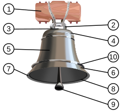
Bell-clapper: 5. Tunica vaginalis. 9. Testes. 

Angular relations between the testes and the tunica vaginalis. Testes lies more horizontal with higher invagination of the tunica.


Cryptochordism is also associated with higher incidence of torsion. Trauma and exercise causing higher cremesteric activity may also result in torsed testes.

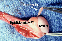
The commoner but less serious torsion of the testicular appendix (Hydatid of Morgagni) occurs also most often at puberty. The appendage is present in 90% of boys. In a review of 771 children with acute scrotum, 58% had torsion of the testicular appendix, 29% of the testes.


Rarely, the testes is torsed between it and the epididymis connected loosely.


Clinical Features

***Epididymo-orchitis is rare in children and adolescents***

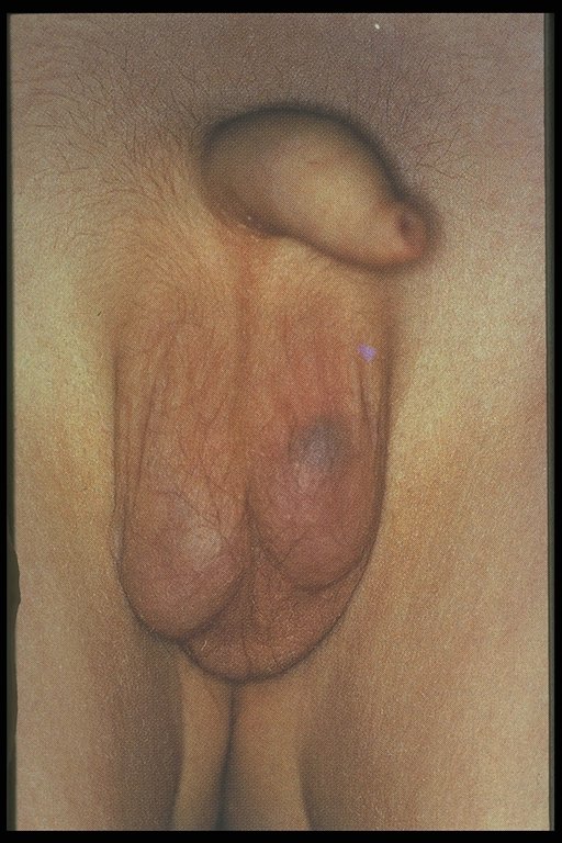
The typical feature is sudden onset of pain at the scrotum or the ipsilateral iliac fossa. This may or may not be accompanied by nausea and vomiting. Beware that some may present with gradual onset of pain leading to delayed diagnosis. Resolving pain may indicate spontaneous resolution of the torsion or worse, a necrotic testes.

Examination reveals inflammed testes which is tender to touch. The testes is usually high riding as well with absence of the cremasteric reflex. However absence of reflex should not be the only indicator as there are case reports of presence of this reflex in a case of testicular torsion. The presence of the bell-clapper anomaly makes the diagnosis more likely and indicated for surgical exploration. Secondary reactive hydrocele may make testicular palpation more difficult.


In a torsed testicular appendix, the pain is usually not as severe and more localized. A 'blue-dot sign' may be seen and point palpation is tender, while palpation of the whole testes is not. However, this is difficult to elicit once secondary hydrocele develop and the scrotum becomes edematous.


Blue-dot sign


Acute scrotum in an infant.


Investigations

***DO NOT LET DELAY IN OBTAINING IMAGING INVESTIGATION DELAY SURGICAL INTERVENTION AND MANAGEMENT***

Ultrasound doppler has a sensitivity of 88% and specificity of 90%. Absence of blood flow to the affected testes is diagnostic. Parenchymal echonegicity is also decreased although it may be increased after infarction. Echogenic, enlarged epididymis is an ancillary sign of testicular torsion. Ultrasound may also help differentiate from hydrocele, abscess, hematoma or tumor.

Torsed appendix usually presents as hyper-echogenic nodule between the epididymis and testes.

Epididymitis may present as hypervascular epididymis although torsion should not be excluded based on just this feature.

Radio-isotope scan may also help in accurately diagnosing testicular torsion. This is unfortunately not widely available.


Note the absence of blood flow in the left testes.


Management

***IF IN DOUBT OF DIAGNOSIS, SURGICALLY EXPLORE***

Manipulation of the testes has been attempted although most suggest that this is done only while waiting for preparation before surgical exploration. This is done in an 'open book' manner, rolling the testes laterally. Success of this maneuver usually relieves the pain spontaneously. This maneuver however is practically difficult to perform on a uncooperative child unless under anesthesia or adequate pain relief. Furthermore, if the torsion occurred in a lateral rotation, the maneuver may worsen the torsion.

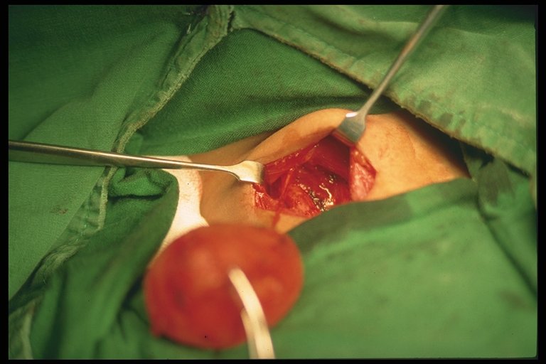
Surgical exploration is warranted if diagnosis is in doubt or there is high suspicion of testicular torsion. Incision may be made paramedian, transverse or vertically through the scrotum. The torsed tested is untwisted.

Examination is done to see testicular viability. If testes is infarcted, orchidopexy is recommended in patients more than 10 years old, as there is risk of autoimmunization with spermatogonia as there is a breech between the blood-testes barrier which may affect fertility at a later age even when the contralateral testes is uninvloved. Some surgeons leave the necrosed testes in-situ in children less than 10 years old.

If the testes is viable, the tested is fixed (orchidopexy). The contralateral testes is also fixed as well, especially when the torsion is associated with the bell-clapper anomaly as this usually occurs bilaterally. It is recommended to use non-absorbable sutures in fixation of the testes tunica albugenia and dartos layer. Use of absorbable sutures are associated with recurrent torsion.


Torsion of the testes. The testes is still viable. Incision of the testes with presence of bleeding may confirm this.


In the torsed appendix of the testes, this is excised.

Necrosed torsioned testicular appendix.


Surgical management is more controversial in torsion in the neonates. 


Prognosis

Rate of testes salvage is inversely proportional with ischemia time. Testes is salvagable by 90% if exploration is performed within 4 - 6 hours after onset of symptoms, 50% if done at 12 hours, and 10% if present more than 24 hours.


Conclusion and Take-Homes

Testicular torsion is a surgical emergency which prompt exploration and treatment is important if the testes were to be salvaged. Diagnosis although may be made by imaging studies, should not cause delay in surgical exploration. The contralateral testes should be explored as well and orchidopexy performed with non-absorbable sutures.


References:

1. Coran, Arnold G., et al., Pediatric Surgery, 7th Ed., Elsevier 

2. Hutson, John M., et al., Jones' Clinical Pediatric Surgery Diagnosis and Management, 6th ed., Blackwell Publishing (2008).

3. Godbole, Prasad P., Testicular Problems in Children, Pediatric and Child Health, Vol 22:6, June 2012.

4. Govindarajan, Krishna K., Pediatric Testicular Torsion, Emedicine, Medscape article.

5. Various websites for the pictures in which none are mine.

Sunday, March 10, 2013

Hirschsprung Disease: Short Notes

Disease caused by absence of ganglions in the myenteric (Auerbach) and submucosal (Meissner) plexus causing functional intestinal obstruction.

Long-segment Hirschsprung: Aganglionic bowel proximal to midtransverse colon 

Embryology and Etiology

Ganglion cells originate from the neural crest tissues by 13 weeks postconception from proximal to distal GIT.

2 theories:
  • Failure of migration of neural crest tissues.
  • Failure of migrated ganglionic cells to mature and proliferate
 Mutation of RET proto-oncogenes

Associated Syndromes

Down syndrome
Neurocristopathy syndromes (ie. Waardenberg-Shah syndrome)
Malrotation
Congenital heart disease
Urinary tract anomalies
Central nervous system anomalies
Congenital central hypoventilation syndrome (Ondine curse)

Clinical Features

Neonates:
  • Delayed passage of meconium after 24 hours
  • Abdominal distension
  • Bilious vomiting
  • Feed intolerance
- Chronic constipation
- Diarrhea, fever, abdominal distension (Hirschprung-associated enterocolitis [HAED])


Investigations

Radiographic
- Abdominal X-ray
- Barium enema

  • Transition zone between normal and aganglionic bowel
  • Reversed recto-sigmoid index (< 1.0)
Anorectal Manometry
  • Recto-anal inhibitory reflex (RAIR). Reflex that causes the internal anal spinchter to relax when the rectum is distended.
  • Balloon inserted into the rectum and inflated. If reflex present detected by sensor.
  • Test more useful with older children.
Rectal biopsy
  • Absence of ganglionic cell
  • Hypertrophied nerve trunk
  • Acetylcholinesterase staining
  • Absence of calcitonin on immunochemical testing with calretinin
Management

Resuscitation
  • Fluids
  • Nasogastric tube
  • Antibiotics
Operative

May be done through laparotomy or more popular, pull-through surgery via the anus.
  • Swenson (end-to-end anastamosis of the ganglionic segment with the internal anal sphincter. Rectum is excised)
  • Soave (As Swenson but rectum is preserved becoming the outer 'cuff' of the pulled ganglinic segment)
  • Duhamel (The ganglinic segment is joined to the rectum in a end-to-side anastamosis)
Colostomy to consider:
  • Enterocolitis
  • Perforated bowel
  • Very dilated proximal bowel
Surgery for Long-segment Hirschsprung:
  • Ileostomy
  • Small bowel pull-through to anastamose with the anal sphincter
  • Side-to-side anastamosis between small bowel and large bowel (to preserve absorptive roles). Several techniques: Martin and Kimura
 Post-op Care
  • Anal calibration 4 to 6 weeks
  • Buttock care with barrier cream to prevent perineal wound breakdown

  
 References:

1. Coran, Arnold G., et al., Pediatric Surgery, 7th Ed., Elsevier

Both pictures from http://radiopaedia.org/cases/hirschsprung-disease 

Saturday, February 16, 2013

Case: Tracheo-esophageal fistula

Baby of RK was born via a Caesarian section for unstable lie at term on the 17th January 2013. AFI was 18. Otherwise no prior anomaly scan was done. She had no other antenatal risk factors. The baby was born vigorous. However at 5 minutes of life noted excessive drooling and the nurse complained of inability to insert the ryles tube deeply during suctioning. The baby had no other physical deformities otherwise.

The child was referred to pediatrics for further evaluation. Chest x-ray revealed the following


Coiling of the ryles tube was noted at the level of the upper chest wall. Stomach air was seen. Vertebrae was normal.

The child was referred to pediatric surgery and an impression of esophageal atresia was made. Surgery was scheduled the next day.

Incision was made below the scapula on the right posterior chest wall up to the anterior axillary line. The chest was opened in layers, the esophagus and trachea exposed via extrapulmonary approach.

Findings of proximal esophageal atresia with distal trache-esophageal fistula was noted. The gap of the defect was about 2 cm. Ligature of the fistula was done followed by primary anastamosis of the esophageal ends with the help of a size 8 Fr ryles tube. Leak test was done after closure. Right chest tube was inserted post-operatively.

Patient was cared in the NICU during which he had complications of left lung atelactasis and worsening of right sided pneumothorax. A new chest tube was inserted reducing the extent of the pneumothorax.

Pneumothorax post-op


Patient also suffered from new onset of sepsis which was treated with antibiotics.

Mechanical ventilation was weaned off, and ryles tube feeding started at day 13 post-op. Breastfeeding initiated at day 24 post-op.

***Discussion to be made later.

Friday, February 15, 2013

Tetralogy of Fallot: The Basics

Pathophysiology:

Tetralogy of Fallot occurs when there is a deviation of the development of the muscular septum of the ventricles antero-cephaladly. This would result in 3 of the 4 features of the 'tetralogy':
  1. Ventricular septal defect (VSD)
  2. Pulmonary stenosis 
  3. Over-riding of the aorta
  4. Right ventricular hypertrophy (Secondary to compensation of the ventricles from pulmonary outflow obstruction)
All these defects occur to variable degrees influencing the manifestation and severity of the condition in affected children.

These defects result in mixing of oxygenated and deoxygenated blood between the VSD from a left-to-right shunt outflow of mixed blood into the aorta causing cyanosis. Cyanosis is worsened when the pulmonary outflow tract is severely restricted and made worse during exacerbation hypercyanotic spell also known as Tet spell.

Compensation may occur via a patent ductus arteriosus (PDA) in newborns and major aortopulmonary collateral arteries (MAPCA).

Clinical Features:
  • Cyanosis. In mild to moderate cases cyanosis may be absent depending on the severity of the pulmonary outflow obstruction.
  • Hypercyanotic spells or Tet spell. 
    • Worsening early in the morning when crying or on exertion or vigorous activities. 
    • May be severe to result in unconsciousness. 
    • Hypoxia may occur to varying degrees which may progress the metabolic acidosis.  
  • Failure to thrive
  • Delayed puberty
  • Clubbing 
  • Parasternal heave
  • Systolic thrill and ejection systolic murmur usually heard over the upper parasternal area due to pulmonay obstruction.
  • Murmur maybe almost absent in hypercyanotic spells due to complete obstruction of the pulmonary outflow tract.
Diagnosis:
  •  Echocardiography: Standard modality for diagnosis. Able to outline extent of defects.
  • Chest X-ray: 
    • Coeur en sabot: Boot shaped heart
    • Right-sided aortic arch
    • Diminished pulmonary vasculature (not prominent)
Boot-shaped heart with right ventricular enlargment.
 
  • ECG:
    • Right axis deviation
    • Prominent R wave on right precordial chest leads and S wave in lateral precordial chest leads.
    • Bifid P wave
Complications:
  • Polycythemia
  • Cerebral thromboses in severe cases of polycythemia
  • Infective endocarditis
  • Brain abscess
ALMOST NEVER HEART FAILURE!

References:

1. Kliegman, Robert M., et al., Nelson Textbook of Pediatrics, 18th ed., Elsevier Saunders.

2. Doyle, T., et al., Pathophysiology, Clinical Features and Diagnosis of Tetralogy of Fallot, UpToDate article, (2013).

3. Bailliard, F., et al, Tetralogy of Fallot.

Future Topics:

- Management of TOF
- Surgical Management of TOF
- Cyanotic heart disease
- Transposition of Great Arteries.

Wednesday, February 13, 2013

Classification of Tracheo-Esophageal Fistula


This classification is anatomically based.

The commonest is still the atretic esophagus with distal fistula as on the left. The middle figure is the isolated Tracheo-esophageal fistula or also known as the 'H' type. Other's are not as common.

Surgical management differs according to anatomical variations.

Other Topics:
- Diagnosis
- Management
- Associated anomalies and VACTERL

Tuesday, February 5, 2013

Kasai Procedure


In 1959, Morio Kasai, a Japanese surgeon discovered a breakthrough in the treatment of biliary atresia which had a poor survival rate before. The procedure which carries his name, although modified in various forms, is still performed today for patient's with biliary atresia.

Procedure


The principle of the procedure is the resect the atretic parts of the extrahepatic biliary tree, dissect the porta hepatis of the liver to free up patent biliary ductules and anastamose with the intestine in a Roux-en-Y fashion (portoenterostomy).

A right upper abdominal incision is made (though from my observation here, the roof top incision is popular). Other anomalies should be sought out and excluded (polysplenia or asplenia, malrotation, preduodenal portal vein, interrupted vena cava).

Before dissection of the portal plate (porta hepatis at the liver surface), examination of the extrahapatic biliary tract is made. If the gallbladder is atretic and without lumen, the surgery may then proceed with the Kasai. If not, an on-table cholangiogram must be performed to delineate the extend of atresia or if atresia exist at all. Failure to outline patent intrahepatic and extrahaptic biliary structures justifies the need to proceed with portoenterostomy. 

The hepatoduodenal ligament (the free edge of the lesser omentum) is opened up to identify the structures of the bile duct and is dissected away from the hepatic arteries. Dissection is continued proximally towards the liver, the gallbladder also dissected. The end-point of dissection is after the bifurcation of the portal vein. A fibrous cone and portal plate should be seen here.

At this stage, the fibrous cone should be transected with knife or scissors. This is the most crucial step to the success of surgery, too deep a cut will cause injury to the liver and cause subsequent scarring and obliteration of the biliary ductules. Too superficial, patent biliary ductules are not exposed enough for drainage. The use of diathermy should be discouraged as this may damage the fine ductules that is essential to the success of the surgery. Care is taken to not injure the portal vein to avoid bleeding.

The proximal jejenum, about 10 cm from the Ligament of Trietze is identified and transected. The distal end of the transection is anatamosed with the liver with 6/0 absorbable sutures brought through the avascular portion of the mesocolon. End-to-side jejunojenunostomy is created at about 50 - 60 cm from the transected part of the jejunum. The defect in the mesocolon is closed by anchoring the roux limb. This is done to prevent internal herniation and keep the limb without tension.

Placement of a suction drain is recommended by some near the portoenterostomy site.

Post-Op

Return of 'color' to stool is an indication of success which usually occur after 10 - 14 days. However some will still progress to liver failure despite initial cholic stool, and some remain with acholic stool.

NG tube is continued until about 48 hours post-op.

Recommended medical regimen includes:
Choleretic: Ursidol : 10 - 15 mg/kg/dose bd
Trimethoprim-sulfamethoxazole (Bactrim) : 2.5 mg/kg/day
Vitamin ADEK
Prednisolone : 2mg/mg/day to be tapered down over 6 weeks.

Factors to Success

Age of surgery seem to be an important factor. Most literature report success if the procedure is performed at 70 to 90 days old. This however does not contraindicate surgery in older children.

Patent gallbladder and fibrous cones are also indicators to better prognosis. So does the diameter of ductules.

Complications

Cholangitis:
An important complication that may lead to the next devastating complication, portal hypertension. Prevention is best done by performing an adequate roux surgery, as well as antibiotics prophylaxis during surgery and post-operatively. The use of steroids also helps in reducing inflammation that may also prevent scarring of the liver although presenting evidence is controversial (not discussed here). Otherwise, treatment is best done with broad-spectrum antibiotics that covers anaerobes as well.

Portal Hypertension:
The usual triad may form including ascites, hypersplenism with associated thrmbocytopenia, and the feared esophageal varices that may lead to bleeding. Liver transplant is indicated if this complication is severe.

Intrahepatic biliary cavities of cyst:
May develop within the liver that may contribute to recurrent cholangitis. Larger ones may be drained percutaneously.

Others:
Wound dehiscence
Internal herniation through the mesocolon defect.
Anastamotic leak
Intussuception at the foot of the roux.

Malignancies:
Cirrhotic liver may lead to hepatocellular carcinoma and hepatoblastoma which all have been reported in patient's with biliary atresia.


References:

1. Coran, Arnold G., et al., Pediatric Surgery, 7th Ed., Elsevier

2. Wildhaber, Barbara E., Biliary Atresia: 50 Years After the First Kasai, Review Article, ISRN Surgery (2012).  Must read. Contains other articles on modified Kasai procedures and others.


Future Topics:

Biliary atresia
Liver Transplant
Complications of biliary atresia (in details)

Monday, February 4, 2013

Use of Negative Pressure Wound therapy for Abdominal Wounds in Neonates and Infants: Critical Appraisal

Use of Negative Pressure Wound Therapy (NPWT) for Abdominal Wounds in Neonates and Infants, Journal of Pediatric Surgery (2012) 47, 1555-1559, Stoffan, Alexander P., et al.

Summary of Article:

The article described the use of NPWT for the past 10 years in Children's Hospital Boston and its outcome. There is no attempts in studying it effectiveness or in comparison to other methods of wound closure or treatment. Only 2 cases developed fistula after initiation of NPWT. Most of the other cases have already developed stoma and fistula before NPWT was applied. 6 died from the cohort but none were related to NPWT.

My 2 cents:

While the article describes the experience of NPWT in their institution, there is a lack useful description to describe the efficacy of NPWT in this age group which is understandable as the article does not intend to report on the subject.

The number of subjects are also too few (18) to be of much benefit.

The wide difference in diagnosis of wounds used for NPWT also makes it difficult to tell if the diagnosis makes a difference to the success of NPWT although some of their results sounds convincing.

Discussion:

Wounds and chronic wounds are a hassle to manage, especially post-operatively. Chronic wounds is a frustrating complications for all parties, including

While not exactly new, its use it being increasingly used in the treatment of especially chronic wounds although any wounds as long as it is not contraindicated, may be treated with this method. In my work in Queen Elizabeth Hospital in Kota Kinabalu, Sabah, its use is increasing especially in the orthopedic's department and general surgery and plastic surgery departments.

The principle to NPWT is that the negative pressure when applied in an air tight seal on a wound, will draw fluids from the wound decreasing edema and encouraging wound healing. It is also said to increase blood flow to the wound which is an important factor in the healing of the wound.

While it is being used in the treatment of wound in adults, its use in the pediatric age group, especially the infant and neonates are also increasing. However, there is a lack of evidence from literature to its efficacy. This is mostly due to the heterogenicity of diagnosis and types of wounds that makes controlled random clinical trials difficult without bias. Despite this, it cannot be denied that NPWT remains an important tool that needs to be utilized.

More research needs to be done surrounding the subject. Wound treatment in the infant and neonate age group may certainly benefit from this modality of treatment. Staff needs to be educated and trained in the application of this technique as it may provide a different alternative when traditional methods of wound care fails.


References:

1. Stoffan, Alexander P., et al., Use of Negative Pressure Wound Therapy (NPWT) for Abdominal Wounds in Neonates and Infants, Journal of Pediatric Surgery (2012) 47, 1555-1559

2. Gestring M, et al., Negative Wound Pressure Therapy, UpToDate Article (2012)

3. Gregor S., et al., Negative Pressure Wound Therapy: A Vacuum of Evidence? Arch Surg 2008

Future Topics:
  • Wound closure methods
  • Wound healing in children

Friday, February 1, 2013

Sistrunk Procedure

Walter Ellis Sistrunk described the surgery for the excision of the thyroglossal duct cyst (TGDC) in 1920.

The principle of the surgery is to remove the cyst along with its tract and the tissues surrounding it including part of the hyoid bone to reduce rate of recurrence of the cyst.

The embryological pathway for the descent of the thyroid gland starting from the foramen caecum, crossing the hyoid bone.


The patient is placed in supine position. A transverse cervical incision is made along the hyoid bone. The cyst and tract is mobilized. A portion of the hyoid bone, about 1 cm each side form the midline is excised after releasing the hyoglossus and mylohyoid muscles. Tissues surrounding the ducts are excised up to the foramen caecum of the tongue. No attempts are made at separating the ducts from the tissue as Sistrunk noted  that the duct are friable and easily broken. The defects are then approximated including the cut hyoid bones.


Cyst with parts of the ducts proximal to it.

On the right is the excised TGDC with a part of the excised hyoid bone and proximal tissues on the left.

Recurrence is about 10% after surgery. Wider excision is recommended for a recurrent cyst. Infected cysts are best drained or treated with antibiotics before proceding with surgery.


References:

1. Sistrunk, W. E., The Surgical Treatment of Cyst of the Thyroglossal Tract, Reprinted from Ann Surg 1920.

2. Coran, Arnold G., et al., Pediatric Surgery, 7th Ed., Elsevier

3. Pictures from various sites as linked.

Other Topics:

- Neck masses in children
- Embryology of the brachial arches

Friday, January 25, 2013

Twin-Twin Transfusion Syndrome: Clinical Features


Twin to twin transfusion syndrome (TTTS) is a condition where there is an over-perfusion in one twin and under-perfusion in the other. Although more common in monochorionic twins, it has been reported to occur in diamniotic twins.

Clinical Features and Diagnosis:

Most important is the discrepancy in amniotic fluid volume.
1 twin would be oligohydramnios ( < 2cm deep vertical pool )
1 twin would be polyhydramnios ( > 8cm deep vertical pool)

WEIGHT DISCREPANCY IS NOT THE MAIN CRITERIA FOR DIAGNOSIS!
In some cases, there are no significant weight discrepancies.
If there are, the discordance should be > 15-20%

"Stuck twin appearance"


Anhydromnios fetus appear as if there are no separating membrane.

Discrepancies in size of umbilical cords.

Presence of hydrops or cardiac dysfunction in recipient twin.

Abnormal umbilical artery doppler in donor fetus.

Other features of monochorionic twins:
  • Single placenta
  • Gender concordance
Fetal Blood Sampling:
- Lower hematocrit level in donors
- Difference in hemoglobin levels.

Staging:

Quintero staging uses simple concept of staging involving presence/absence of oligo or polyhydromnios, bladder, abnormal dopple, hydrops fetalis, or fetal death.

A table can be seen in this Emedicine website.

References:

Creasy, Robert K., et al., Maternal-Fetal Medicine, Principles and Practice, 5th Ed.

Other Topics To Discuss:

- Management of TTTS
- Monochorionic twins
- Conjoint twins.