Saturday, February 16, 2013

Case: Tracheo-esophageal fistula

Baby of RK was born via a Caesarian section for unstable lie at term on the 17th January 2013. AFI was 18. Otherwise no prior anomaly scan was done. She had no other antenatal risk factors. The baby was born vigorous. However at 5 minutes of life noted excessive drooling and the nurse complained of inability to insert the ryles tube deeply during suctioning. The baby had no other physical deformities otherwise.

The child was referred to pediatrics for further evaluation. Chest x-ray revealed the following


Coiling of the ryles tube was noted at the level of the upper chest wall. Stomach air was seen. Vertebrae was normal.

The child was referred to pediatric surgery and an impression of esophageal atresia was made. Surgery was scheduled the next day.

Incision was made below the scapula on the right posterior chest wall up to the anterior axillary line. The chest was opened in layers, the esophagus and trachea exposed via extrapulmonary approach.

Findings of proximal esophageal atresia with distal trache-esophageal fistula was noted. The gap of the defect was about 2 cm. Ligature of the fistula was done followed by primary anastamosis of the esophageal ends with the help of a size 8 Fr ryles tube. Leak test was done after closure. Right chest tube was inserted post-operatively.

Patient was cared in the NICU during which he had complications of left lung atelactasis and worsening of right sided pneumothorax. A new chest tube was inserted reducing the extent of the pneumothorax.

Pneumothorax post-op


Patient also suffered from new onset of sepsis which was treated with antibiotics.

Mechanical ventilation was weaned off, and ryles tube feeding started at day 13 post-op. Breastfeeding initiated at day 24 post-op.

***Discussion to be made later.

Friday, February 15, 2013

Tetralogy of Fallot: The Basics

Pathophysiology:

Tetralogy of Fallot occurs when there is a deviation of the development of the muscular septum of the ventricles antero-cephaladly. This would result in 3 of the 4 features of the 'tetralogy':
  1. Ventricular septal defect (VSD)
  2. Pulmonary stenosis 
  3. Over-riding of the aorta
  4. Right ventricular hypertrophy (Secondary to compensation of the ventricles from pulmonary outflow obstruction)
All these defects occur to variable degrees influencing the manifestation and severity of the condition in affected children.

These defects result in mixing of oxygenated and deoxygenated blood between the VSD from a left-to-right shunt outflow of mixed blood into the aorta causing cyanosis. Cyanosis is worsened when the pulmonary outflow tract is severely restricted and made worse during exacerbation hypercyanotic spell also known as Tet spell.

Compensation may occur via a patent ductus arteriosus (PDA) in newborns and major aortopulmonary collateral arteries (MAPCA).

Clinical Features:
  • Cyanosis. In mild to moderate cases cyanosis may be absent depending on the severity of the pulmonary outflow obstruction.
  • Hypercyanotic spells or Tet spell. 
    • Worsening early in the morning when crying or on exertion or vigorous activities. 
    • May be severe to result in unconsciousness. 
    • Hypoxia may occur to varying degrees which may progress the metabolic acidosis.  
  • Failure to thrive
  • Delayed puberty
  • Clubbing 
  • Parasternal heave
  • Systolic thrill and ejection systolic murmur usually heard over the upper parasternal area due to pulmonay obstruction.
  • Murmur maybe almost absent in hypercyanotic spells due to complete obstruction of the pulmonary outflow tract.
Diagnosis:
  •  Echocardiography: Standard modality for diagnosis. Able to outline extent of defects.
  • Chest X-ray: 
    • Coeur en sabot: Boot shaped heart
    • Right-sided aortic arch
    • Diminished pulmonary vasculature (not prominent)
Boot-shaped heart with right ventricular enlargment.
 
  • ECG:
    • Right axis deviation
    • Prominent R wave on right precordial chest leads and S wave in lateral precordial chest leads.
    • Bifid P wave
Complications:
  • Polycythemia
  • Cerebral thromboses in severe cases of polycythemia
  • Infective endocarditis
  • Brain abscess
ALMOST NEVER HEART FAILURE!

References:

1. Kliegman, Robert M., et al., Nelson Textbook of Pediatrics, 18th ed., Elsevier Saunders.

2. Doyle, T., et al., Pathophysiology, Clinical Features and Diagnosis of Tetralogy of Fallot, UpToDate article, (2013).

3. Bailliard, F., et al, Tetralogy of Fallot.

Future Topics:

- Management of TOF
- Surgical Management of TOF
- Cyanotic heart disease
- Transposition of Great Arteries.

Wednesday, February 13, 2013

Classification of Tracheo-Esophageal Fistula


This classification is anatomically based.

The commonest is still the atretic esophagus with distal fistula as on the left. The middle figure is the isolated Tracheo-esophageal fistula or also known as the 'H' type. Other's are not as common.

Surgical management differs according to anatomical variations.

Other Topics:
- Diagnosis
- Management
- Associated anomalies and VACTERL

Tuesday, February 5, 2013

Kasai Procedure


In 1959, Morio Kasai, a Japanese surgeon discovered a breakthrough in the treatment of biliary atresia which had a poor survival rate before. The procedure which carries his name, although modified in various forms, is still performed today for patient's with biliary atresia.

Procedure


The principle of the procedure is the resect the atretic parts of the extrahepatic biliary tree, dissect the porta hepatis of the liver to free up patent biliary ductules and anastamose with the intestine in a Roux-en-Y fashion (portoenterostomy).

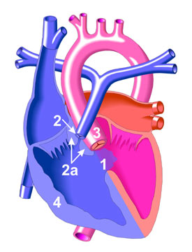
A right upper abdominal incision is made (though from my observation here, the roof top incision is popular). Other anomalies should be sought out and excluded (polysplenia or asplenia, malrotation, preduodenal portal vein, interrupted vena cava).

Before dissection of the portal plate (porta hepatis at the liver surface), examination of the extrahapatic biliary tract is made. If the gallbladder is atretic and without lumen, the surgery may then proceed with the Kasai. If not, an on-table cholangiogram must be performed to delineate the extend of atresia or if atresia exist at all. Failure to outline patent intrahepatic and extrahaptic biliary structures justifies the need to proceed with portoenterostomy. 

The hepatoduodenal ligament (the free edge of the lesser omentum) is opened up to identify the structures of the bile duct and is dissected away from the hepatic arteries. Dissection is continued proximally towards the liver, the gallbladder also dissected. The end-point of dissection is after the bifurcation of the portal vein. A fibrous cone and portal plate should be seen here.

At this stage, the fibrous cone should be transected with knife or scissors. This is the most crucial step to the success of surgery, too deep a cut will cause injury to the liver and cause subsequent scarring and obliteration of the biliary ductules. Too superficial, patent biliary ductules are not exposed enough for drainage. The use of diathermy should be discouraged as this may damage the fine ductules that is essential to the success of the surgery. Care is taken to not injure the portal vein to avoid bleeding.

The proximal jejenum, about 10 cm from the Ligament of Trietze is identified and transected. The distal end of the transection is anatamosed with the liver with 6/0 absorbable sutures brought through the avascular portion of the mesocolon. End-to-side jejunojenunostomy is created at about 50 - 60 cm from the transected part of the jejunum. The defect in the mesocolon is closed by anchoring the roux limb. This is done to prevent internal herniation and keep the limb without tension.

Placement of a suction drain is recommended by some near the portoenterostomy site.

Post-Op

Return of 'color' to stool is an indication of success which usually occur after 10 - 14 days. However some will still progress to liver failure despite initial cholic stool, and some remain with acholic stool.

NG tube is continued until about 48 hours post-op.

Recommended medical regimen includes:
Choleretic: Ursidol : 10 - 15 mg/kg/dose bd
Trimethoprim-sulfamethoxazole (Bactrim) : 2.5 mg/kg/day
Vitamin ADEK
Prednisolone : 2mg/mg/day to be tapered down over 6 weeks.

Factors to Success

Age of surgery seem to be an important factor. Most literature report success if the procedure is performed at 70 to 90 days old. This however does not contraindicate surgery in older children.

Patent gallbladder and fibrous cones are also indicators to better prognosis. So does the diameter of ductules.

Complications

Cholangitis:
An important complication that may lead to the next devastating complication, portal hypertension. Prevention is best done by performing an adequate roux surgery, as well as antibiotics prophylaxis during surgery and post-operatively. The use of steroids also helps in reducing inflammation that may also prevent scarring of the liver although presenting evidence is controversial (not discussed here). Otherwise, treatment is best done with broad-spectrum antibiotics that covers anaerobes as well.

Portal Hypertension:
The usual triad may form including ascites, hypersplenism with associated thrmbocytopenia, and the feared esophageal varices that may lead to bleeding. Liver transplant is indicated if this complication is severe.

Intrahepatic biliary cavities of cyst:
May develop within the liver that may contribute to recurrent cholangitis. Larger ones may be drained percutaneously.

Others:
Wound dehiscence
Internal herniation through the mesocolon defect.
Anastamotic leak
Intussuception at the foot of the roux.

Malignancies:
Cirrhotic liver may lead to hepatocellular carcinoma and hepatoblastoma which all have been reported in patient's with biliary atresia.


References:

1. Coran, Arnold G., et al., Pediatric Surgery, 7th Ed., Elsevier

2. Wildhaber, Barbara E., Biliary Atresia: 50 Years After the First Kasai, Review Article, ISRN Surgery (2012).  Must read. Contains other articles on modified Kasai procedures and others.


Future Topics:

Biliary atresia
Liver Transplant
Complications of biliary atresia (in details)

Monday, February 4, 2013

Use of Negative Pressure Wound therapy for Abdominal Wounds in Neonates and Infants: Critical Appraisal

Use of Negative Pressure Wound Therapy (NPWT) for Abdominal Wounds in Neonates and Infants, Journal of Pediatric Surgery (2012) 47, 1555-1559, Stoffan, Alexander P., et al.

Summary of Article:

The article described the use of NPWT for the past 10 years in Children's Hospital Boston and its outcome. There is no attempts in studying it effectiveness or in comparison to other methods of wound closure or treatment. Only 2 cases developed fistula after initiation of NPWT. Most of the other cases have already developed stoma and fistula before NPWT was applied. 6 died from the cohort but none were related to NPWT.

My 2 cents:

While the article describes the experience of NPWT in their institution, there is a lack useful description to describe the efficacy of NPWT in this age group which is understandable as the article does not intend to report on the subject.

The number of subjects are also too few (18) to be of much benefit.

The wide difference in diagnosis of wounds used for NPWT also makes it difficult to tell if the diagnosis makes a difference to the success of NPWT although some of their results sounds convincing.

Discussion:

Wounds and chronic wounds are a hassle to manage, especially post-operatively. Chronic wounds is a frustrating complications for all parties, including

While not exactly new, its use it being increasingly used in the treatment of especially chronic wounds although any wounds as long as it is not contraindicated, may be treated with this method. In my work in Queen Elizabeth Hospital in Kota Kinabalu, Sabah, its use is increasing especially in the orthopedic's department and general surgery and plastic surgery departments.

The principle to NPWT is that the negative pressure when applied in an air tight seal on a wound, will draw fluids from the wound decreasing edema and encouraging wound healing. It is also said to increase blood flow to the wound which is an important factor in the healing of the wound.

While it is being used in the treatment of wound in adults, its use in the pediatric age group, especially the infant and neonates are also increasing. However, there is a lack of evidence from literature to its efficacy. This is mostly due to the heterogenicity of diagnosis and types of wounds that makes controlled random clinical trials difficult without bias. Despite this, it cannot be denied that NPWT remains an important tool that needs to be utilized.

More research needs to be done surrounding the subject. Wound treatment in the infant and neonate age group may certainly benefit from this modality of treatment. Staff needs to be educated and trained in the application of this technique as it may provide a different alternative when traditional methods of wound care fails.


References:

1. Stoffan, Alexander P., et al., Use of Negative Pressure Wound Therapy (NPWT) for Abdominal Wounds in Neonates and Infants, Journal of Pediatric Surgery (2012) 47, 1555-1559

2. Gestring M, et al., Negative Wound Pressure Therapy, UpToDate Article (2012)

3. Gregor S., et al., Negative Pressure Wound Therapy: A Vacuum of Evidence? Arch Surg 2008

Future Topics:
  • Wound closure methods
  • Wound healing in children

Friday, February 1, 2013

Sistrunk Procedure

Walter Ellis Sistrunk described the surgery for the excision of the thyroglossal duct cyst (TGDC) in 1920.

The principle of the surgery is to remove the cyst along with its tract and the tissues surrounding it including part of the hyoid bone to reduce rate of recurrence of the cyst.

The embryological pathway for the descent of the thyroid gland starting from the foramen caecum, crossing the hyoid bone.


The patient is placed in supine position. A transverse cervical incision is made along the hyoid bone. The cyst and tract is mobilized. A portion of the hyoid bone, about 1 cm each side form the midline is excised after releasing the hyoglossus and mylohyoid muscles. Tissues surrounding the ducts are excised up to the foramen caecum of the tongue. No attempts are made at separating the ducts from the tissue as Sistrunk noted  that the duct are friable and easily broken. The defects are then approximated including the cut hyoid bones.


Cyst with parts of the ducts proximal to it.

On the right is the excised TGDC with a part of the excised hyoid bone and proximal tissues on the left.

Recurrence is about 10% after surgery. Wider excision is recommended for a recurrent cyst. Infected cysts are best drained or treated with antibiotics before proceding with surgery.


References:

1. Sistrunk, W. E., The Surgical Treatment of Cyst of the Thyroglossal Tract, Reprinted from Ann Surg 1920.

2. Coran, Arnold G., et al., Pediatric Surgery, 7th Ed., Elsevier

3. Pictures from various sites as linked.

Other Topics:

- Neck masses in children
- Embryology of the brachial arches